For decades, Alzheimer’s disease has been one of the hardest brain disorders to treat, largely because it is usually diagnosed only after memory loss and confusion disrupt everyday life. By then, extensive damage has already occurred in the…
Category: 6. Health
-
In surprising breakthrough, scientists in Israel find cancer may help heal the failing heart
A discovery by Prof. Ami Aronheim and his team at the Technion–Israel Institute of Technology shows that the growth of cancerous tumors may actually combat cardiac dysfunction and reduce fibrosis, the scarring process that stiffens the…
Continue Reading
-
Gut microbiota-derived metabolite isovalerylcarnitine modulates salt sensitivity of blood pressure and incident hypertension: a multicenter dietary salt intervention trial
Global Burden of Disease. Global burden of disease https://vizhub.healthdata.org/gbd-compare/ (2022).
Wang, Y. J., Yeh, T. L., Shih, M. C., Tu, Y. K. & Chien, K. L. Dietary sodium intake and risk of cardiovascular disease: a systematic review and…
Continue Reading
-
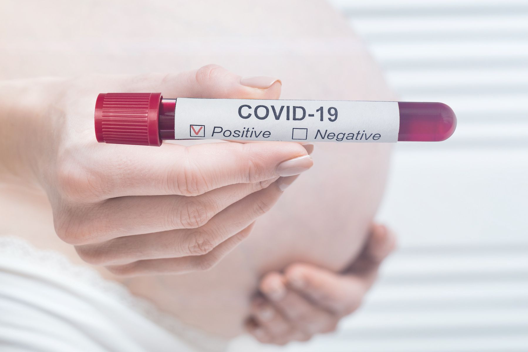
COVID-19 Vaccination Is Associated With Lower Risk of Maternal Disease
Immunization against the SARS-CoV-2 virus prior to or during pregnancy yielded a lower risk of severe maternal disease and preterm birth, according to a study published in JAMA.1 Although variants of the virus were known to impact various…
Continue Reading
-

Eating more vitamin C can physically change your skin
Scientists at the University of Otago, Faculty of Medicine — Christchurch Ōtautahi, have identified a direct connection between how much vitamin C people eat and how well their skin produces collagen and renews itself. The findings show that…
Continue Reading
-

The perfect morning routine: how to build a happy, healthy start to the day – from showers to sunshine | Health & wellbeing
The first thing to say about the ideal morning routine is that it probably doesn’t exist. Yes, endless influencers promise that they have tweaked, tested and fine-tuned the process of revving up for the day, but how history’s most productive…
Continue Reading
-
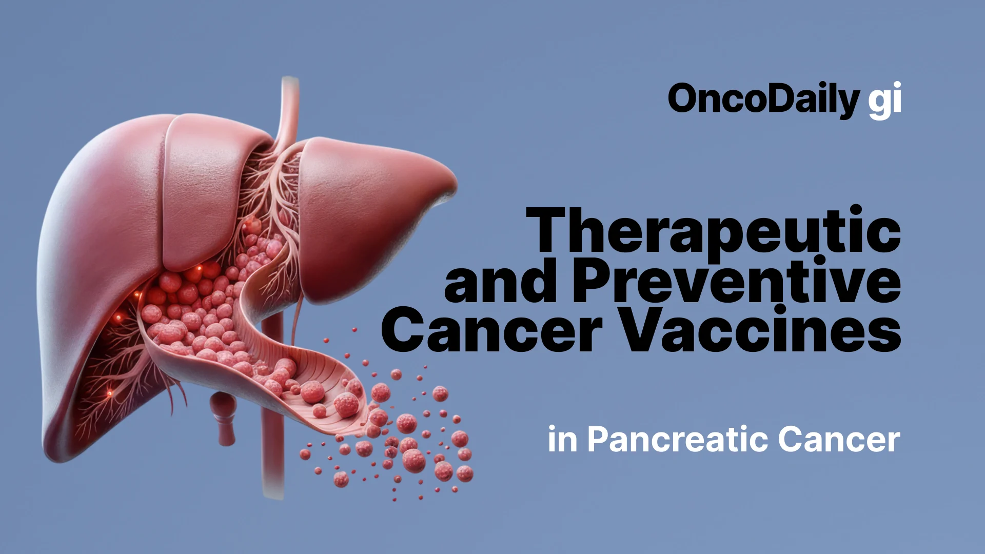
Therapeutic Vaccines in Pancreatic Cancer: Challenging a Historically Immune-Resistant Disease
Pancreatic cancer remains one of the most lethal malignancies worldwide, with five-year survival rates hovering around 10% and median survival often measured in months rather than years. The majority of patients present with…
Continue Reading
-

Stent Bridging vs Emergency Surgery in Obstructive Colon Cancer
A bridge-to-surgery approach using colonic stent placement offers similar short-term survival outcomes to emergency surgery in patients with left-sided obstructive colorectal cancer, while reducing the need for stomas and enabling more…
Continue Reading
-

Pediatric surgeons weigh AI benefits against ethical challenges and practical barriers
Artificial intelligence (AI) is rapidly advancing across modern healthcare, yet its role in pediatric surgery remains limited and ethically complex. This study reveals that although surgeons recognize AI’s potential to enhance…
Continue Reading