Aleix Prat, Director of the Clínic Barcelona Comprehensive Cancer Center at Barcelona Clinical Hospital, shared a post on LinkedIn:
“2025 has been a very special year for HER2DX
Looking back, it’s been quite striking to see…
Aleix Prat, Director of the Clínic Barcelona Comprehensive Cancer Center at Barcelona Clinical Hospital, shared a post on LinkedIn:
“2025 has been a very special year for HER2DX
Looking back, it’s been quite striking to see…

Depression and anxiety are independently associated with an increased risk of major adverse cardiovascular events (MACE) and are partially mediated by heightened stress-related neural activity, highlighting shared stress-related pathophysiology…

Malaria remains one of the deadliest infectious diseases, which is responsible for approximately 249 million cases and over 600,000 deaths globally every year, with children under 5 in sub-Saharan Africa bearing the greatest burden. The recent…

Runcie C.W. Chidebe/LinkedIn
Runcie C.W. Chidebe, Executive Director at Project PINK BLUE, shared a post on LinkedIn about a recent article he and his colleagues co-authored,…
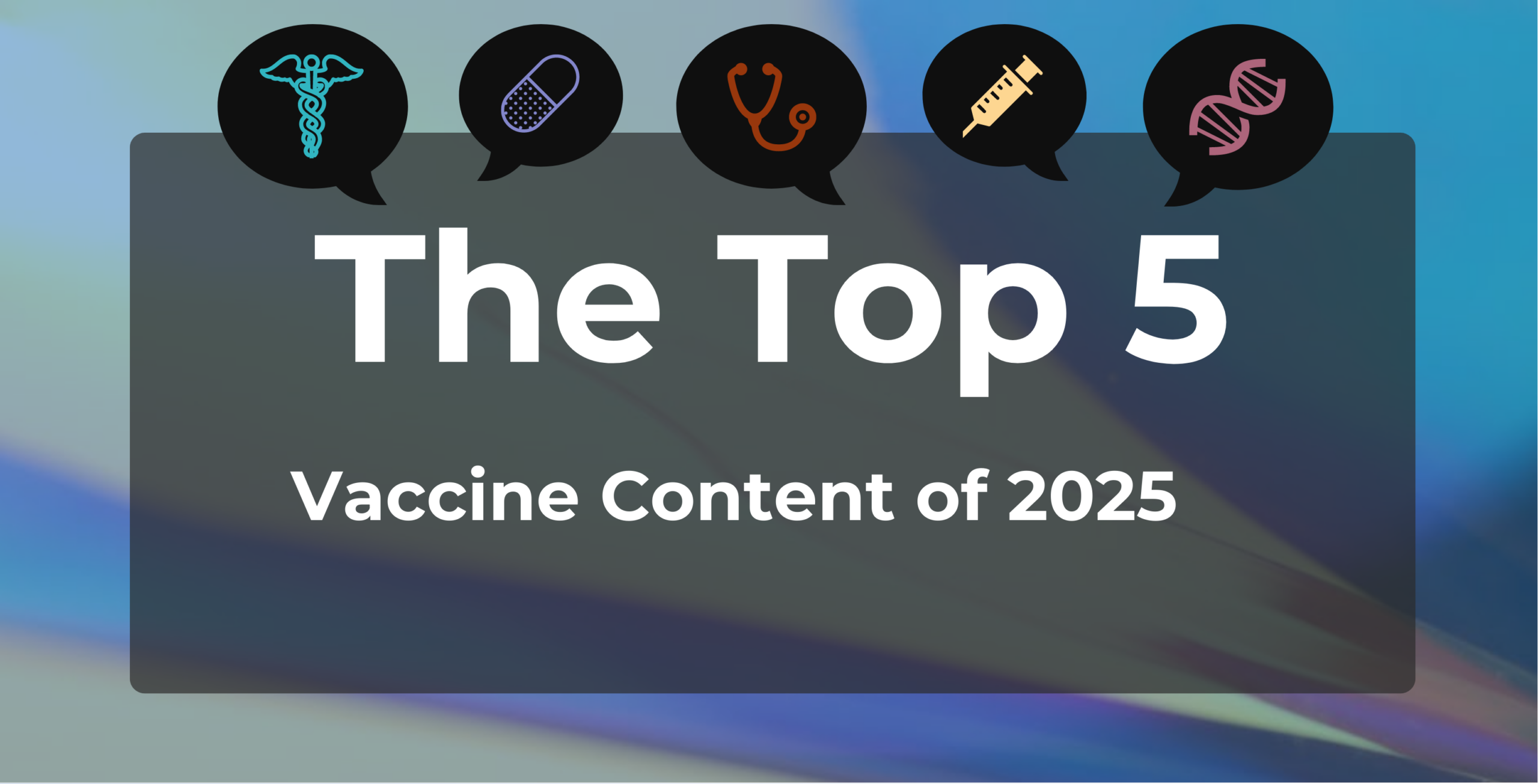
In 2025, several developments converged to shake public and professional confidence in US vaccination policy. Advisory panels were overhauled, former leaders sounded alarms, lawsuits emerged, and new guidelines were poised to overturn…
Mittal, R., Rath, S. & Vemuganti, G. K. Ocular surface squamous neoplasia – Review of etio-pathogenesis and an update on clinico-pathological diagnosis. Saudi J. Ophthalmol. 27, 177–186. https://doi.org/10.1016/J.SJOPT.2013.07.002 (2013).

It was another busy year for podcasts on CancerNetwork®, as 2025 saw continued collaboration with experts in several different specialties to provide critical in-depth perspectives from all corners of the oncology world. Between the community…
Stroke, or cerebrovascular accident (CVA), is the second leading cause of death and the third leading cause of death and disability combined among non-communicable diseases (NCDs) globally, with China having the highest stroke…

With everyday advancement, new…