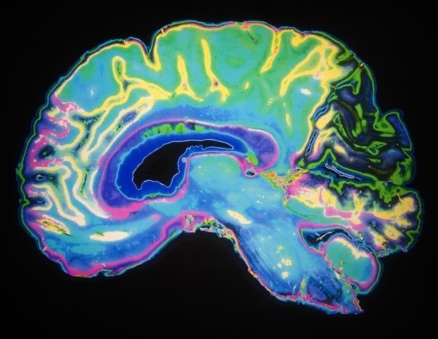
Siemens Healthineers announced today its offerings for brain health research are expanding, with the brain-derived, fully automated Atellica IM Phosphorylated tau 217 (pTau217) and Atellica IM Brain Derived Tau (BDTau) assays now…
Category: 6. Health
-
Peng Liyuan calls for global action against TB
BEIJING – Peng Liyuan, wife of Chinese President Xi Jinping and also the World Health Organization (WHO) goodwill ambassador for tuberculosis (TB) and HIV/AIDS, on Wednesday called for international support and participation in global TB…
Continue Reading
-

Menopause may raise women’s Alzheimer risk earlier than doctors once thought
A new review suggests the menopause transition may mark a critical window for Alzheimer’s prevention in women, shifting focus toward earlier detection, sex-specific risk factors, and more personalized care.
Expert Review: Women’s…
Continue Reading
-
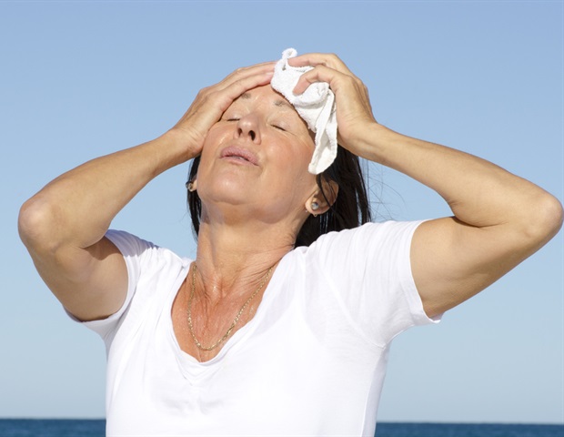
Early menopause linked to higher lifetime heart disease risk
Women who enter natural menopause before age 40 face about a 40% higher lifetime risk of developing coronary heart disease than women who experience menopause later, according to a large Northwestern Medicine study that is the first…
Continue Reading
-
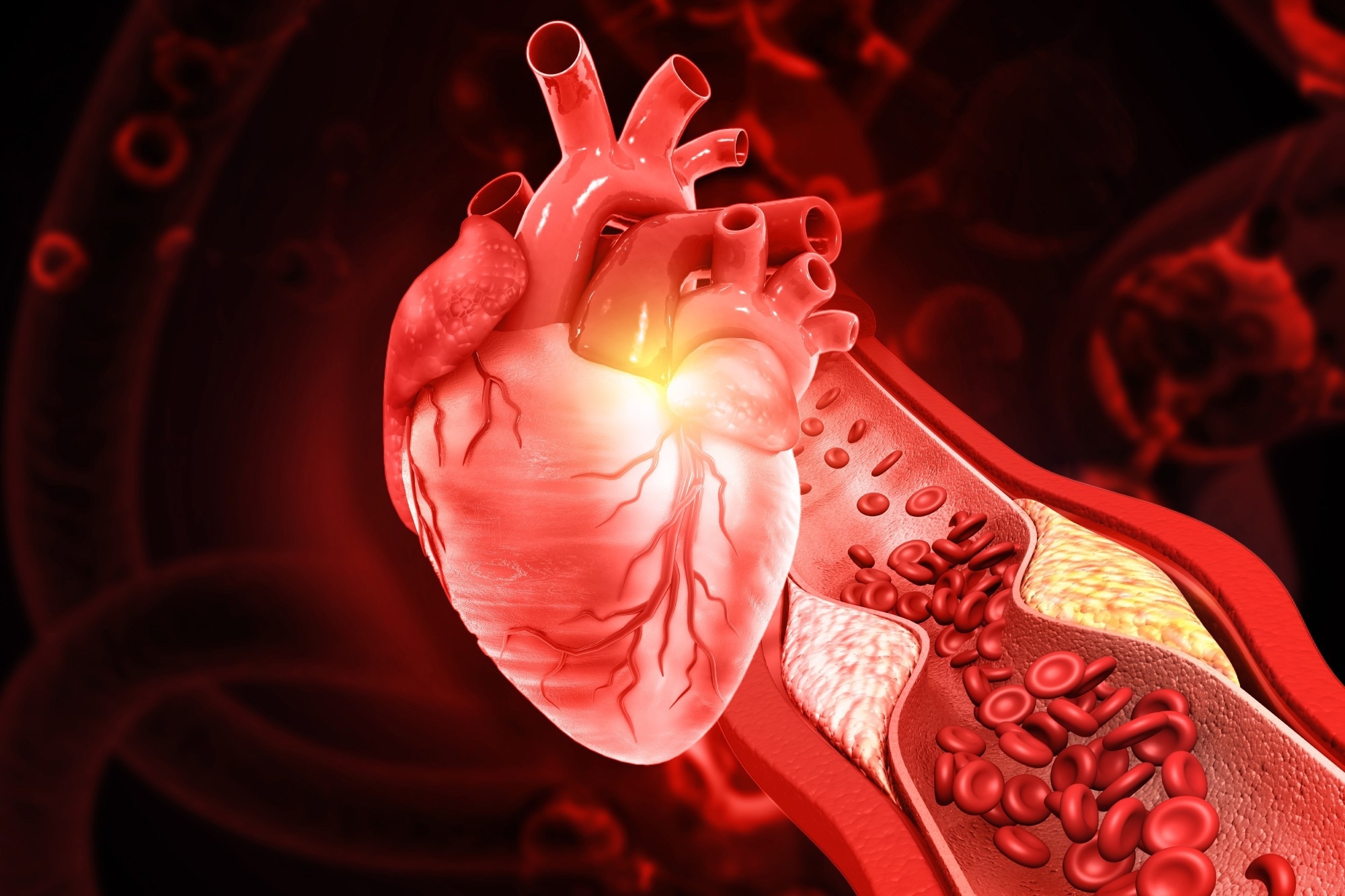
Gut-derived blood markers may help predict who develops coronary heart disease
A major multi-cohort study found that several gut microbiota-related metabolites in the bloodstream were linked to later coronary heart disease, pointing to new biomarker and therapeutic targets while underscoring that the evidence…
Continue Reading
-

The surprising cancer link between cats and humans
The first large-scale analysis of multiple cancer types in cats has uncovered genetic changes that may help guide new treatments for both animals and people.
Researchers examined tumors from nearly 500 pet cats across five countries. The work…
Continue Reading
-

This simple habit could help seniors live longer and stay independent
In Japan, many older adults rely on bicycles for daily transportation, far more than seniors in Europe or the United States. Earlier research has shown that people who cycle tend to be more physically active and socially engaged. While cycling…
Continue Reading
-
Rabies in Argentina: The past 5 years
During the period spanning 2021 to 2026, 10 suspected cases of human rabies were investigated IN Argentina; of these, one was a confirmed case, recorded in the province of Buenos Aires in 2021. The remaining cases yielded negative laboratory…
Continue Reading
-
Brain Aging Tied to MIND Diet Scores – MedPage Today
- Brain Aging Tied to MIND Diet Scores MedPage Today
- Brain aging slowed by over 2 years with unique diet CNN
- 3 Neurologists Share Their Go-To Food for Cognitive Health EatingWell
- Healthy eating may help keep the brain younger, study suggests
Continue Reading
-
Beyond seizures: With experimental drug, Stoke aims to restore developmental loss in Dravet syndrome – Fierce Biotech
- Beyond seizures: With experimental drug, Stoke aims to restore developmental loss in Dravet syndrome Fierce Biotech
- Why Stoke Therapeutics (STOK) Is Down 11.4% After NEJM Data on Dravet Syndrome Drug – And What’s Next simplywall.st
- National…
Continue Reading