Throughout the past year, NeurologyLive® served as a platform not only for interviews and expert commentary, but also for original clinician authored perspectives that addressed some of the most pressing and evolving issues in neurology….
Category: 6. Health
-

EBV Antibodies Linked to Autoantibody Levels in Rheumatoid Arthritis
New findings suggest that immune responses to Epstein–Barr virus (EBV) may be associated with heightened autoimmunity in rheumatoid arthritis (RA). In a retrospective observational study, patients with RA who tested positive for anti-EBV…
Continue Reading
-

Free Skin Anterolateral Thigh Flap And Vacuum-Assisted Closure Therapy
Introduction
Snakebite envenomation constitutes a significant public health burden in tropical nations, particularly in Vietnam, where bites from cobra (Naja species) are prevalent.1,2 While systemic toxicity from cobra bites may be mild, the…
Continue Reading
-

Blood test could predict who is most at risk from common inherited heart condition | Health
Scientists are developing a simple blood test to predict who is most at risk from the world’s most common inherited heart condition.
Millions of people worldwide have hypertrophic cardiomyopathy (HCM), a disease of the heart muscle where the…
Continue Reading
-
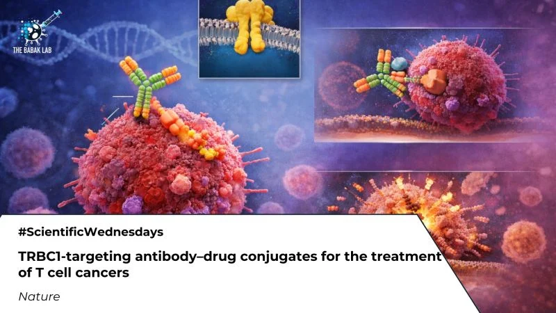
Scientific Wednesdays: TRBC1-Targeting ADCs for T-Cell Malignancies – The Babak Lab
The Babak Lab shared a post on LinkedIn:
“Scientific Wednesdays: TRBC1-Targeting ADCs for T-Cell Malignancies
A new Nature study introduces TRBC1-targeting antibody-drug conjugates (ADCs) as a precision strategy for treating…
Continue Reading
-
Preliminary Developmental Safety Assessment of Allylestrenol in Pregna
Introduction
Progesterone is a key hormone that plays an essential role in the maintenance of pregnancy.1 In early gestation, it supports embryo implantation and uterine quiescence by reducing tubal contractility, modulating cervical mucus,…
Continue Reading
-
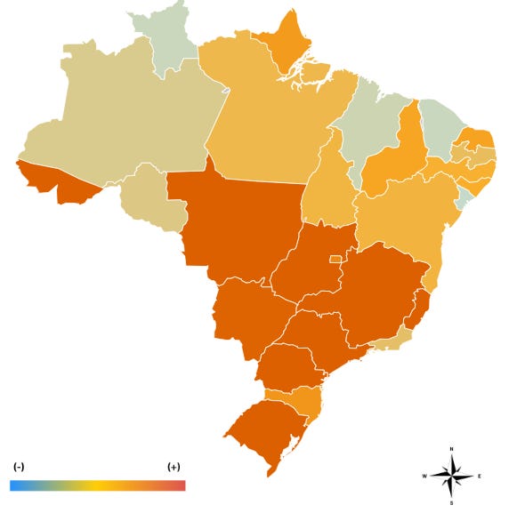
Brazil dengue cases down 75% in 2025
Brazil recorded 1,660,190 probable cases of dengue fever in 2025, according to the most recent update from the Ministry of Health. During the same period, the disease caused 1,762 deaths, while another 200 deaths are still under investigation….
Continue Reading
-
Atypical hemolytic uremic syndrome after post-abortion infection: case
Introduction
Thrombotic microangiopathy (TMA) is characterized by organ failure, microangiopathic hemolytic anemia, and thrombocytopenia. It is particularly relevant to the brain (unconsciousness, seizures), kidneys (acute renal injury), and…
Continue Reading
-
Exploring Theoretical Models Used to Explain Factors Influencing Breas
Introduction
Breast cancer (BC) represents the most common cancer among women in 86% of countries.1 In 2022, BC mortality was ranked fourth worldwide, as reported by the International Agency for Research on Cancer.2 Annually, around 1.7 million…
Continue Reading
-

Gianrico Farrugia: Emily’s story—a 2030 patient experience
Gianrico Farrugia, President and CEO at Mayo Clinic, shared a post on LinkedIn:
“In this post, we follow a patient, Emily, on a transformed healthcare journey.
Mayo Clinic is leading patient-centered healthcare transformation…
Continue Reading