Category: 6. Health
-
Health officials confirm case of measles in Central Virginia and more headlines – Virginia Mercury
- Health officials confirm case of measles in Central Virginia and more headlines Virginia Mercury
- A person with measles traveled in Virginia and North Carolina, officials say The Washington Post
- Virginia Health officials investigating potential…
Continue Reading
-

Breakfast tips to keep energy levels steady for the winters
Whether you’re at risk for type 2 diabetes or just trying to keep your energy levels…
Continue Reading
-

Oral Bacteria and Alzheimer’s Disease May Intersect – European Medical Journal Oral Bacteria and Alzheimer’s Disease Risk
ORAL bacteria and Alzheimer’s disease may be linked as periodontitis inflammation could promote neurodegeneration, authors suggest.
Oral Bacteria and Alzheimer’s Disease: What the Evidence Shows
A new editorial highlights a growing body of…
Continue Reading
-

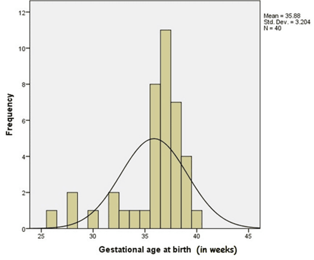
A study on the correlation between Triglyceride-glucose index (TyG ind
Introduction
Coronary artery disease (CAD) is a leading cause of mortality worldwide, characterized by myocardial ischemia and hypoxia due to coronary artery constriction or obstruction.1 Early and accurate diagnosis of CAD is essential, with…
Continue Reading
-

Biomarkers And Genetics In Heart Risk Prediction
NEW evidence suggests cardiovascular risk prediction can be significantly improved by combining clinical biomarkers, metabolomics and genetics, potentially allowing clinicians to identify high risk individuals earlier and prevent more…
Continue Reading
-
Find out if you have it
It’s estimated that around 982,000 people in the UK are living with dementia,…
Continue Reading
-
Your browser is not supported
Your browser is not supported | southcoasttoday.com 
southcoasttoday.com wants to ensure the best experience for all of our readers, so we built our site to take advantage of the latest technology, making it faster and easier to use.
Unfortunately,…
Continue Reading
-
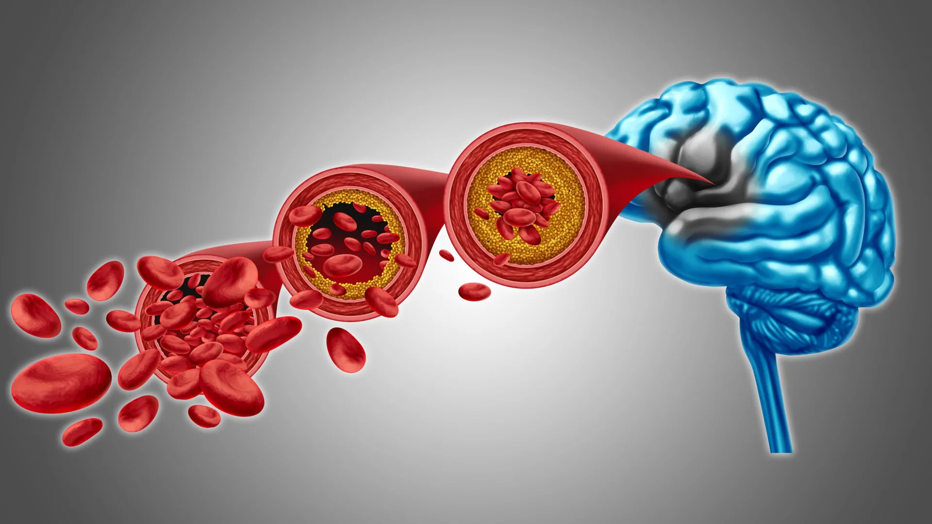
Scientists found a way to restore brain blood flow in dementia
A potential new way to treat reduced brain blood flow and certain forms of dementia is beginning to emerge. Scientists at the University of Vermont Robert Larner, M.D. College of Medicine have uncovered new details about how blood circulation in…
Continue Reading
-

New drug treatment set to change ways of alcoholism
An 18-week experimental study, published in Alcohol Clinical & Experimental Research, examining…
Continue Reading