eople, the holidays often bring joyful indulgence, followed by regret and ambitious New Year’s resolutions to eat better.
A recent study from the University of British Columbia suggests moderation should not be a seasonal goal but a long-term…

A recent study from the University of British Columbia suggests moderation should not be a seasonal goal but a long-term…
A study reveals that restoring the brain’s energy balance may not just slow Alzheimer’s — but actually reverse it.
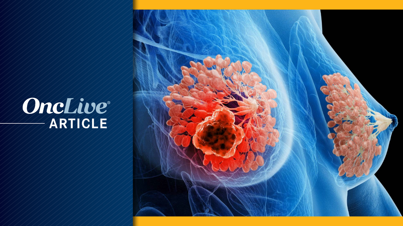
The therapeutic landscape for hormone receptor–positive, HER2-negative breast cancer is currently undergoing a paradigm shift, characterized by the expansion of oral selective estrogen receptor degraders (SERDs) and targeted therapies into…

Editor’s note: Welcome to a Science and Culture Today tradition: a countdown of our Top 10 favorite stories of the past year, concluding on New Year’s Day. This article was originally published on October 9, 2025. Our staff are…

The amount of flu circulating has started to fall in England, latest data suggests.
The UK Health Security Agency (UKHSA) said it was encouraging news heading into Christmas, but warned the virus could always bounce back in the new year.
The UKHSA…
Estrogen, a protective sex hormone, is widely recognized for its beneficial effects on cardiovascular health. This leads to sex differences in cardiovascular health: men have a higher incidence of CVD, whereas women experience…
Presubicular layer 4 neurons are intrinsic bursting pyramidal neurons that project to the lateral mammillary nucleus (Huang et al., 2017). This pathway is critical for the coordinated stable landmark control of HD cells in the thalamus and…

Refugee health is often discussed in terms of crises such as disease outbreaks, malnutrition and psychological distress. But some of the most serious effects of displacement are harder to see. One example is how forced migration can change…
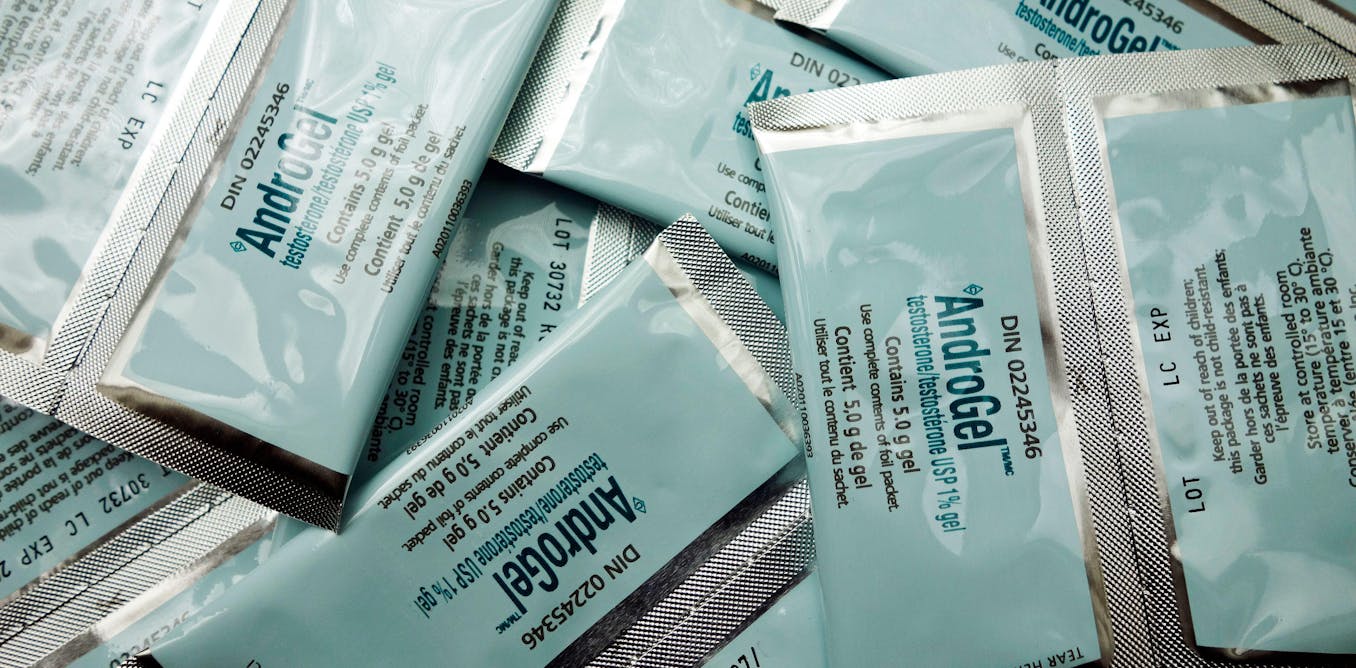
For more than 80 years, men have been told that testosterone helps prostate cancer grow. But a very different picture has emerged over the past two decades.
The prostate is a small gland that sits just below the bladder. Its job is to…
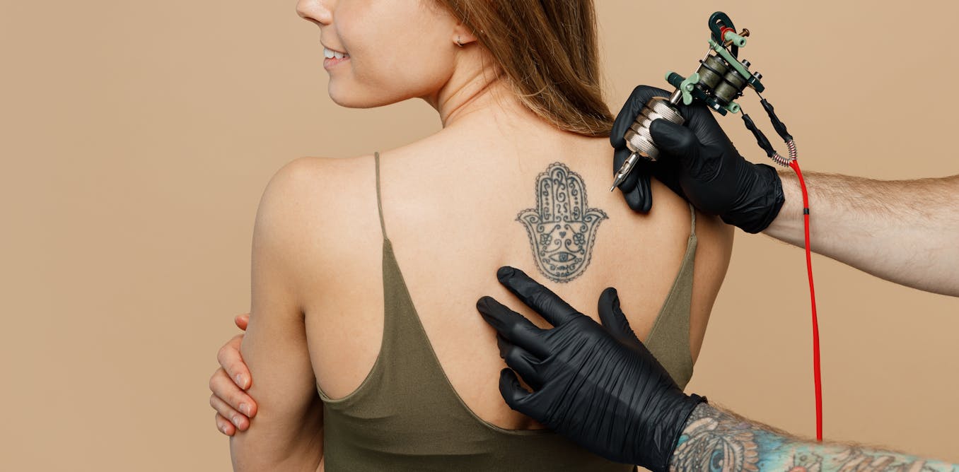
From minimalist wrist designs to full sleeves, body art has become so common that it barely raises an eyebrow. But while the personal meaning of a tattoo may be obvious, the biological consequences are far less visible. Once tattoo ink…