A new observational study in the Journal of Clinical Oncology – Oncology Practice provides real-world evidence demonstrating feasibility of 5-fluorouracil, leucovorin, oxaliplatin, and irinotecan (FOLFIRINOX; FFX) chemotherapy in carefully…
Category: 6. Health
-
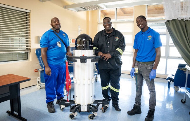
How Trinidad and Tobago is reimagining healthcare through innovation – PAHO/WHO
Just one click with perinatal digital health records
If robots and fridges have strengthened Trinidad’s infrastructure, the country’s application of the Perinatal Information System (SIP and SIP Plus) is strengthening information.
Digitizing…
Continue Reading
-
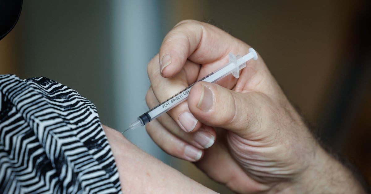
Flu cases among people aged 65 and older up by almost 25% over past week – The Irish Times
Cases of flu among people aged 65 and older have increased by almost 25 per cent over the last week, new figures show.
On Tuesday, the Health Protection Surveillance Centre (HPSC) published the latest figures for respiratory illnesses –…
Continue Reading
-

Distribution features and trends of antimicrobial resistance patterns
Introduction
Antimicrobial resistance (AMR) is increasingly acknowledged as a big global danger to health in the 21st century. A systematic analysis of AMR from 1990 to 2021, with forecasts extending to 2050, underscores the escalating threat…
Continue Reading
-

Beyond Access: The Hidden Cultural Barriers to Cancer Screening Across South Asia, the Middle East, and Africa
Early cancer screening is an important part of cancer control strategies. It significantly contributes to reduced mortality through timely detection and treatment of malignancies. However, despite medical advantages, screening…
Continue Reading
-

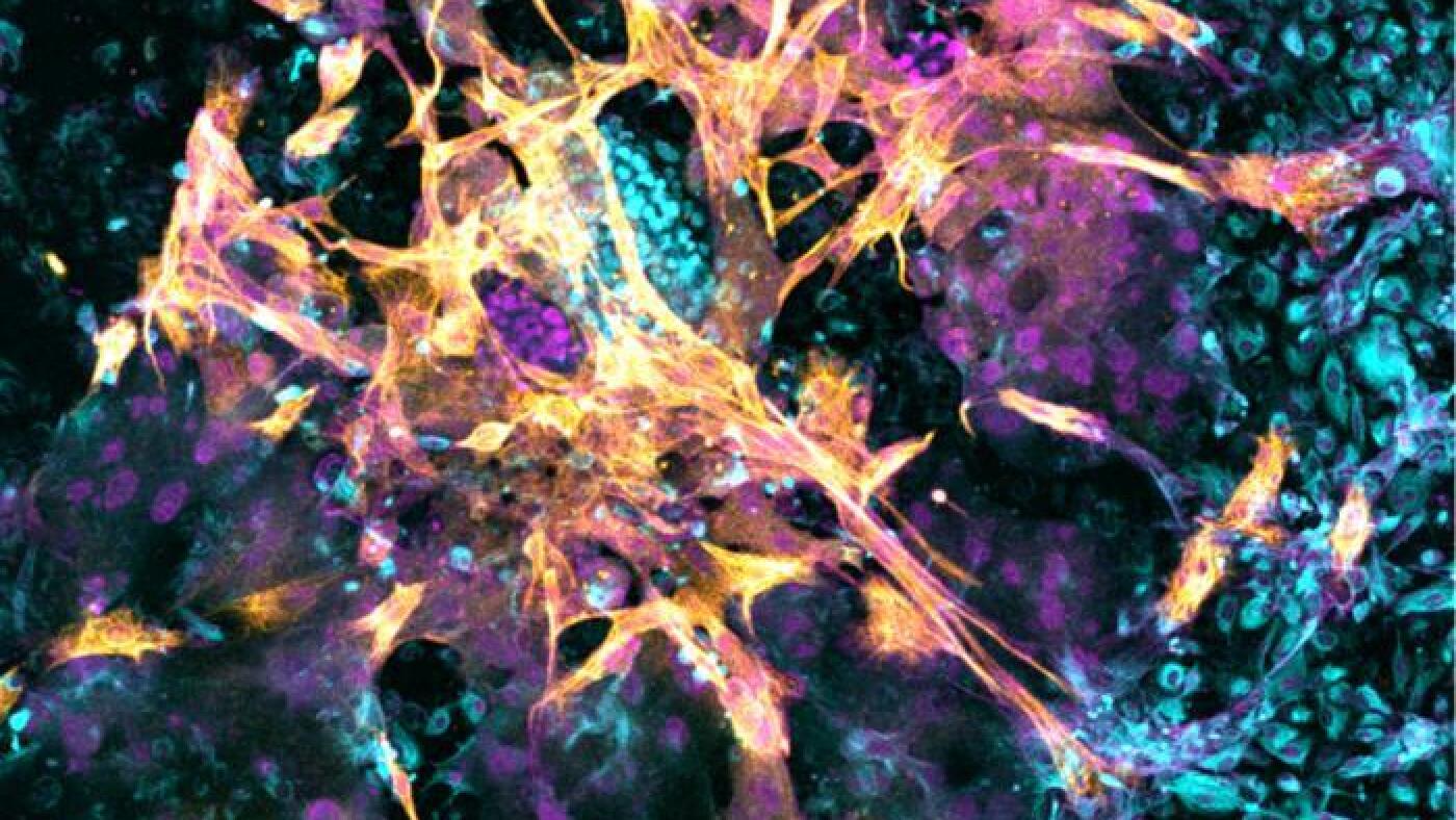
Scientists create replica human womb lining and implant early-stage embryos | Reproduction
Researchers have created the lining of a womb in a dish, which promises to shed light on the mysterious early stages of human pregnancy and the glitches that can lead to miscarriage and medical complications.
In laboratory experiments, early-stage…
Continue Reading
-
Scientists learn more about how human embryos implant using artificial wombs : NPR
Microscopy image of a day 14 human embryo that has implanted in the new artificial womb.
…Continue Reading
-
Residence, education ,motivation for fluid intake,autonomous perceptio
Introduction
Maintenance hemodialysis (MHD) serves as a life-sustaining transitional therapy for patients with end-stage renal disease (ESRD) and remains the primary treatment modality currently available for ESRD management.1 Hemodialysis…
Continue Reading
-
Clinical Comparison of Group A Streptococcus Antigen Detection and Pat
Introduction
Group A Streptococcus (GAS) infection is a common bacterial disease in children, often causing upper respiratory tract infections such as pharyngitis and tonsillitis, and, in severe cases, leading to complications such as rheumatic…
Continue Reading
-
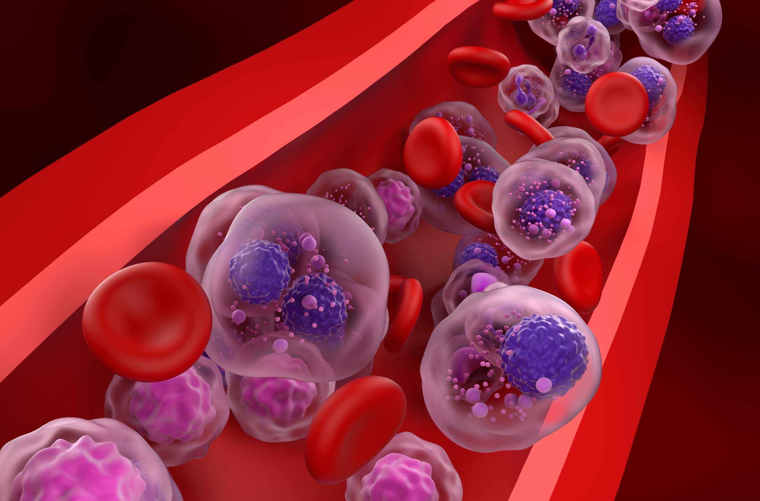
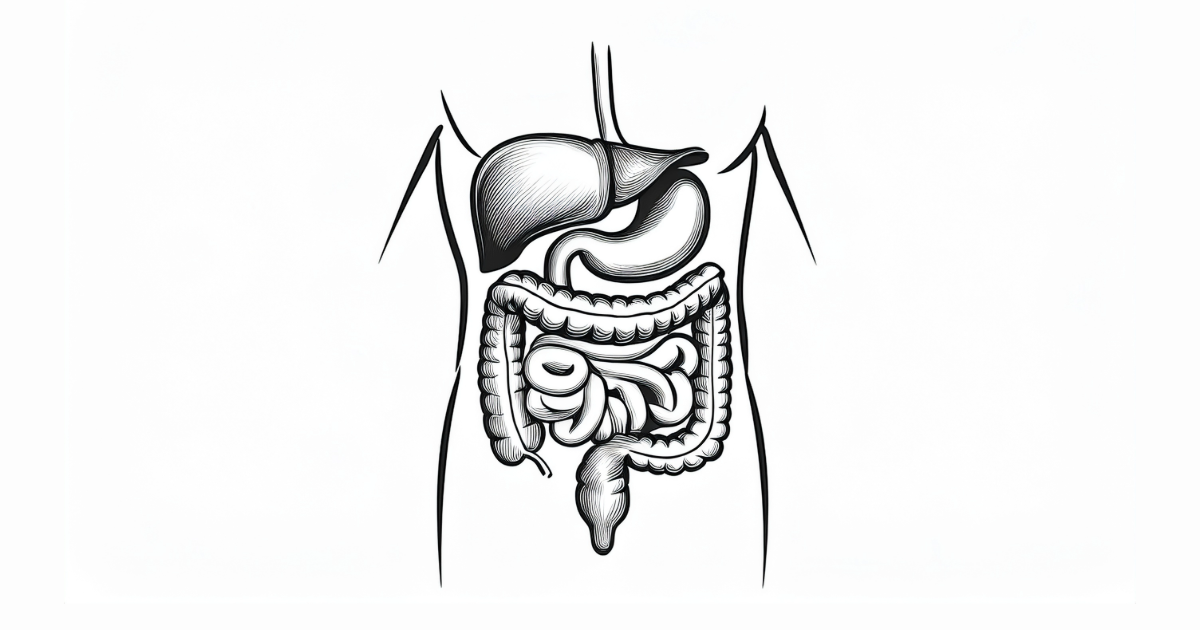
High-Fiber Diet May Delay Progression of Multiple Myeloma in Patients With MGUS, SMM
Research published in Cancer Discovery suggests that a high-fiber, plant-based diet delayed progression of multiple myeloma (MM) in patients with monoclonal gammopathy of undetermined significance (MGUS) and smoldering multiple myeloma (SMM)….
Continue Reading