Category: 6. Health
-
Reviewing ‘Aging in Wellness and Adversity,’ on dementia
Reviewing ‘Aging in Wellness and Adversity,’ on dementia | The Jerusalem Post -
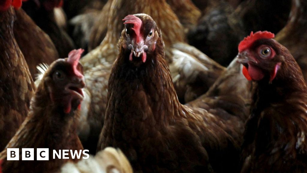
Two more cases of bird flu at Dereham poultry sites
Two more cases of bird flu have been confirmed at large commercial poultry sites in Norfolk.
The Department for Environment, Food and Rural Affairs (Defra) said the H5N1 virus was confirmed at two premises near Dereham, in Breckland, on 8 December…
Continue Reading
-
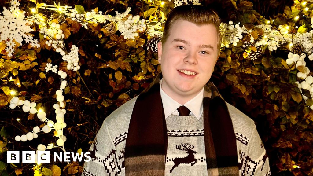
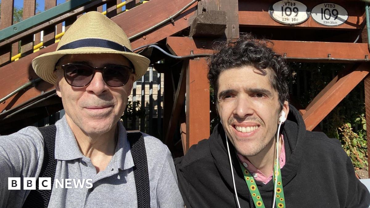
Bury boy, 17, releases charity Christmas album after ME diagnosis
 Harry Boulton
Harry Boulton, 17, has released a charity single of White Christmas to help others with chronic fatigue system A teenager has released a charity Christmas album after being diagnosed with ME (myalgic encephalomyelitis), also known as…
Continue Reading
-
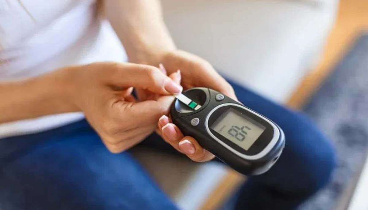
Researchers uncover breakthrough drug for diabetes
Scientists at NYU Langone Health have discovered a new drug compound that may help treat some…
Continue Reading
-
Redhill father’s appeal after son’s brain tumour death
 Family handout
Chavier de Abreu (right) died from a brain tumour in April 2024 A father facing his second Christmas without his son, who died from a brain tumour, is supporting an appeal to help fund research into the disease.
Chavier de Abreu, 34,…
Continue Reading
-
Redhill father’s appeal after son’s brain tumour death
A father facing his second Christmas without his son, who died from a brain tumour, is supporting an appeal to help fund research into the disease.
Chavier de Abreu, 34, who was known as Chevy, died from an astrocytoma in 2024, three years after…
Continue Reading
-

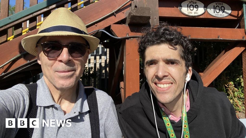
Home at Christmas for Newcastle boy, 8, given cancer all-clear
Jaxen’s treatment left him neutropenic, which meant his body was low in white blood cells that protect against infection.
During previous Christmases, he was only allowed one visitor in hospital, because of the risk of infection.
Jaxen said he…
Continue Reading
-
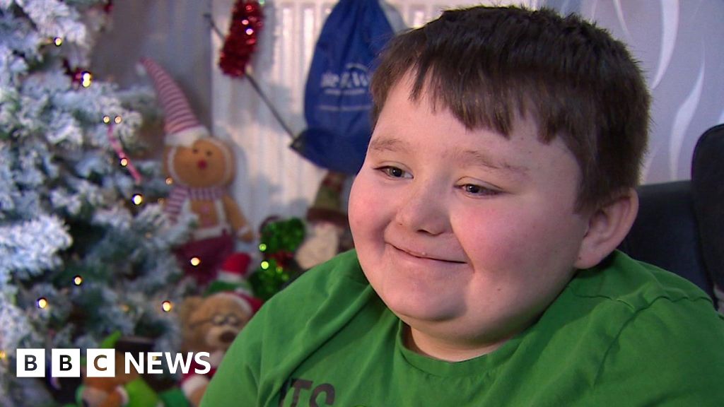
Home at Christmas for Newcastle boy, 8, given cancer all-clear
Helen RichardsonNorth East and Cumbria
 BBC
BBCEight-year-old Jaxen will be at home this Christmas after three years of cancer treatment A young boy will spend Christmas at home after getting the all-clear from cancer.
Eight-year-old Jaxen, from…
Continue Reading
-

Body awareness is fundamental to longevity, according to an expert trainer—here’s how to improve yours
For most of us, the way to increase your chances of living for longer in good health is pretty straightforward.
Strength training, cardio work and flexibility routines can all improve your longevity, but according to trainer Eloise Skinner,…
Continue Reading
-

Brain tumour teenager gets message of support from Harry Maguire
England and Manchester United footballer Harry Maguire has sent a message to a 14-year-old who was diagnosed with a brain tumour at the end of last month.
Max from Corby, Northamptonshire, went into hospital just six days after his birthday and…
Continue Reading