England and Manchester United footballer Harry Maguire has sent a message to a 14-year-old who was diagnosed with a brain tumour at the end of last month.
Max from Corby, Northamptonshire, went into hospital just six days after his birthday and…
England and Manchester United footballer Harry Maguire has sent a message to a 14-year-old who was diagnosed with a brain tumour at the end of last month.
Max from Corby, Northamptonshire, went into hospital just six days after his birthday and…
England and Manchester United footballer Harry Maguire has sent a message to a 14-year-old who was diagnosed with a brain tumour at the end of last month.
Max from Corby, Northamptonshire, went into hospital just six days after his birthday and…
We’ve all…

A relatively mundane middle-aged rite of passage — shingles vaccination — might offer an added benefit: protection against or even the slowing of the progression of dementia.
Over the past several years,…

PUBLISHED
December 21, 2025
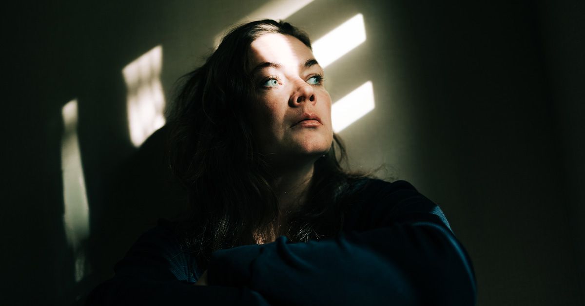
Inmates serving their sentences look forward to the day…

Despite having voluminous, wavy-curly hair that often leads people to assume I have great density, there are days when I gather it into a ponytail and sense something is off – it feels thinner, lighter, and less substantial than it used to. For…

Willamette Week is in the middle of our most important annual fundraiser. As a local independent news outlet, we need your help.
Give today. Hold power to account.
This story was produced by the Oregon Journalism Project, a nonprofit newsroom…