
Two years into an emotionally draining mission to get pregnant, with no sign of a positive result, Idaho couple Darby and Jesse Nubbe were feeling desperate. “We were $16,000 (£12,000) out of pocket, with weekly blood work, invasive…
Category: 6. Health
-
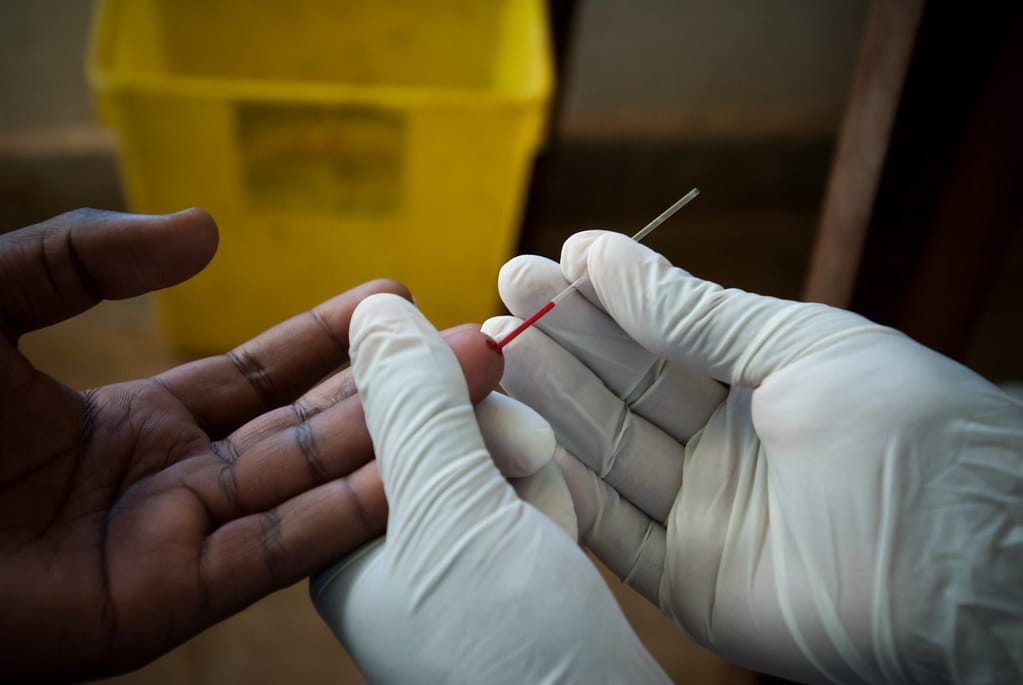
Younger, mobile men especially likely to not know they have HIV in eastern and southern Africa
aidsmap news story
One in seven men living with HIV in eastern and southern Africa are unaware that they have the virus, according to research presented at CROI 2026. Younger men and those who sometimes live away from home were more likely not to…
Continue Reading
-
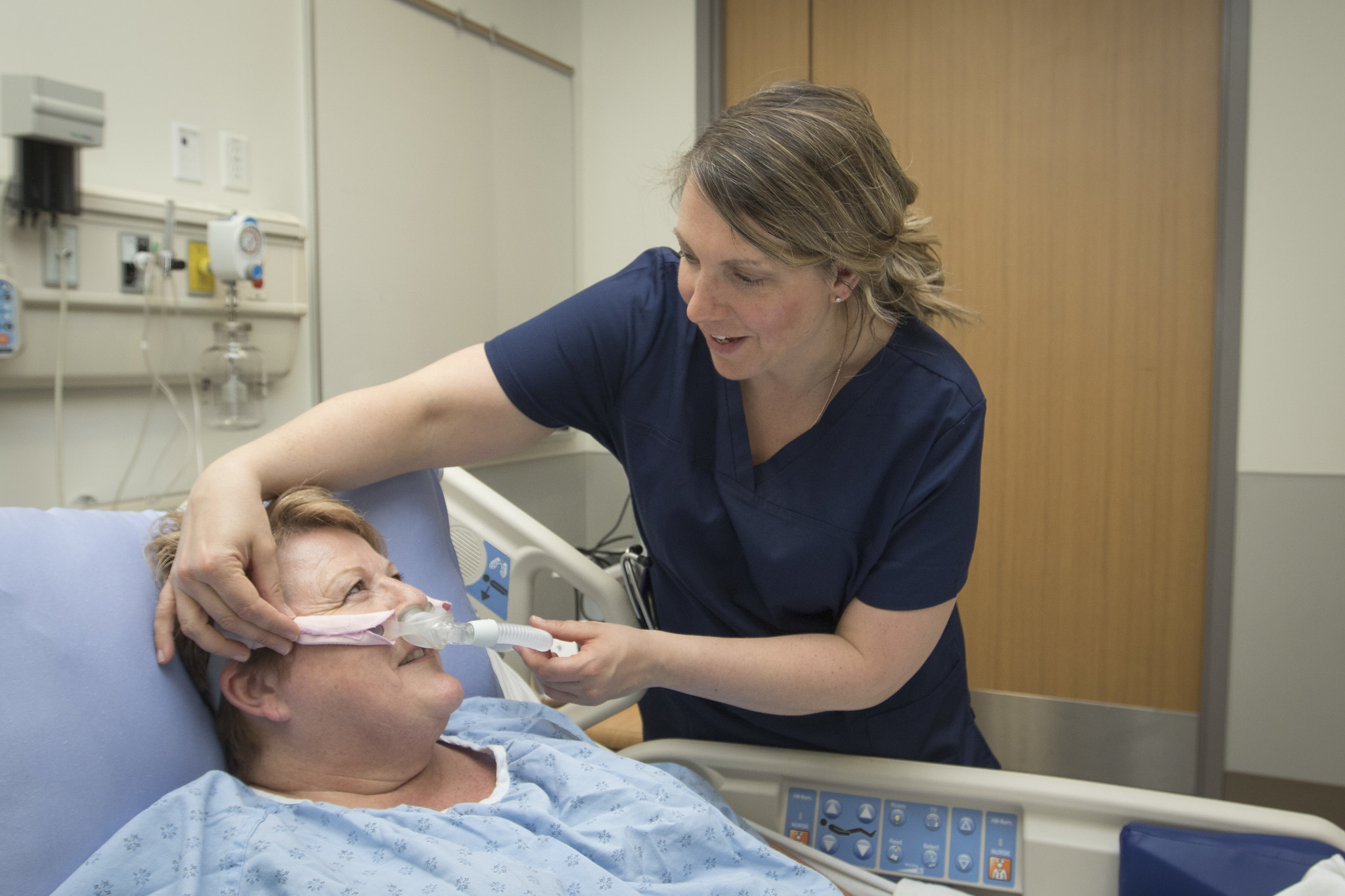
Study Reveals Promising Pill for Obstructive Sleep Apnea Treatment
A new experimental pill may offer hope to millions of people living with obstructive sleep apnea, a condition that repeatedly disrupts breathing during sleep. Researchers in Europe report that the drug significantly reduced breathing…
Continue Reading
-
Six-week virtual program offers early palliative care roadmap for dementia – Medical Xpress
- Six-week virtual program offers early palliative care roadmap for dementia Medical Xpress
- A roadmap for dementia care: Early palliative care offers support for patients and caregivers facing dementia The Medical University of South Carolina
Continue Reading
-
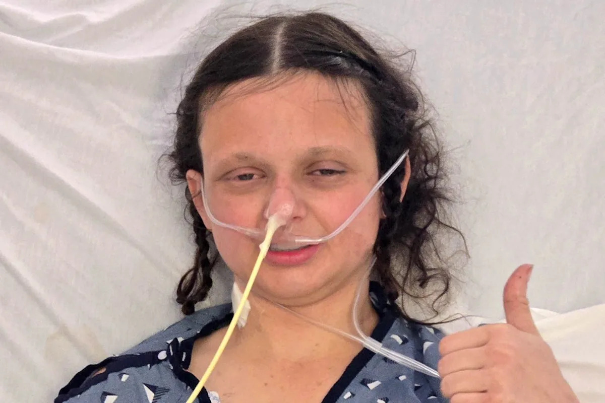
2-Time Cancer Survivor, 24, Facing Life-Threatening Illness After She Ate Friend’s Homemade Meal
NEED TO KNOW
-
Trinity Peterson-Mayes, 24, is facing a life-threatening illness after eating a homemade meal with her friends
-
“I was intubated, I had a central line in my neck, and I had an NG [nasogastric] tube… and I just woke up and I couldn’t…
Continue Reading
-
-

Study Findings: Obesity linked to higher risk of severe infections
Adult obesity significantly increases the risk of severe infections and infection-related deaths, according to a large international study published in The Lancet. The research suggests that around one in ten infection-related deaths globally may…
Continue Reading
-

Your body’s “biological stress score” may reveal disease risk years early
This strain does not come from one event. It builds slowly from sleep loss, diet, work pressure, sedentary habits, and even environmental exposure. Over time, these pressures affect the heart, metabolism, immune system, and brain.
Doctors…
Continue Reading
-
Weekly Health Update | Namibia Ends Mpox Outbreak; Nigeria Faces Seasonal Lassa Fever Surge
This week, Africa is facing a mixed health situation. Namibia has declared an end to its mpox outbreak, while Madagascar is reporting rising case counts. Nigeria is grappling with a seasonal surge in Lassa fever. The World Health…
Continue Reading
-

GLP-1RAs Could Help Type 2 Diabetes and Brain Cancer
PATIENTS with brain metastases and Type 2 diabetes using glucagon-like peptide-1 receptor agonists (GLP-1RA) have a near 40% lower risk of death over 3 years compared with nonusers, a 2026 retrospective cohort study has found.
Comorbid Type 2…
Continue Reading
-

Social connections may be key to older adults staying physically active
A strong social network can encourage older adults to be more physically active – leading to better health and mental well-being, researchers say.
A team from the Texas A&M University School of Public Health reviewed 34 research articles,…
Continue Reading