 Getty Images
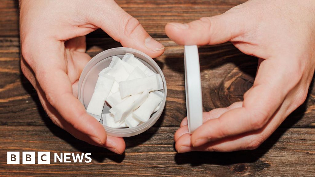
Young men are behind the growing popularity of nicotine pouches in Great Britain, a study suggests.
Around 7.5% of 16 to 24-year-old-men are using the small sachets that fit under the top lip, compared with 1.9% use among young women…
 Getty Images
Young men are behind the growing popularity of nicotine pouches in Great Britain, a study suggests.
Around 7.5% of 16 to 24-year-old-men are using the small sachets that fit under the top lip, compared with 1.9% use among young women…

New WellSpace Provides Unique and Innovative Way of Promoting Staff and Physician Wellness
Rady Children’s Health has opened its first “Associate WellSpace,” a biophilic, restorative space designed to support the emotional and physical…

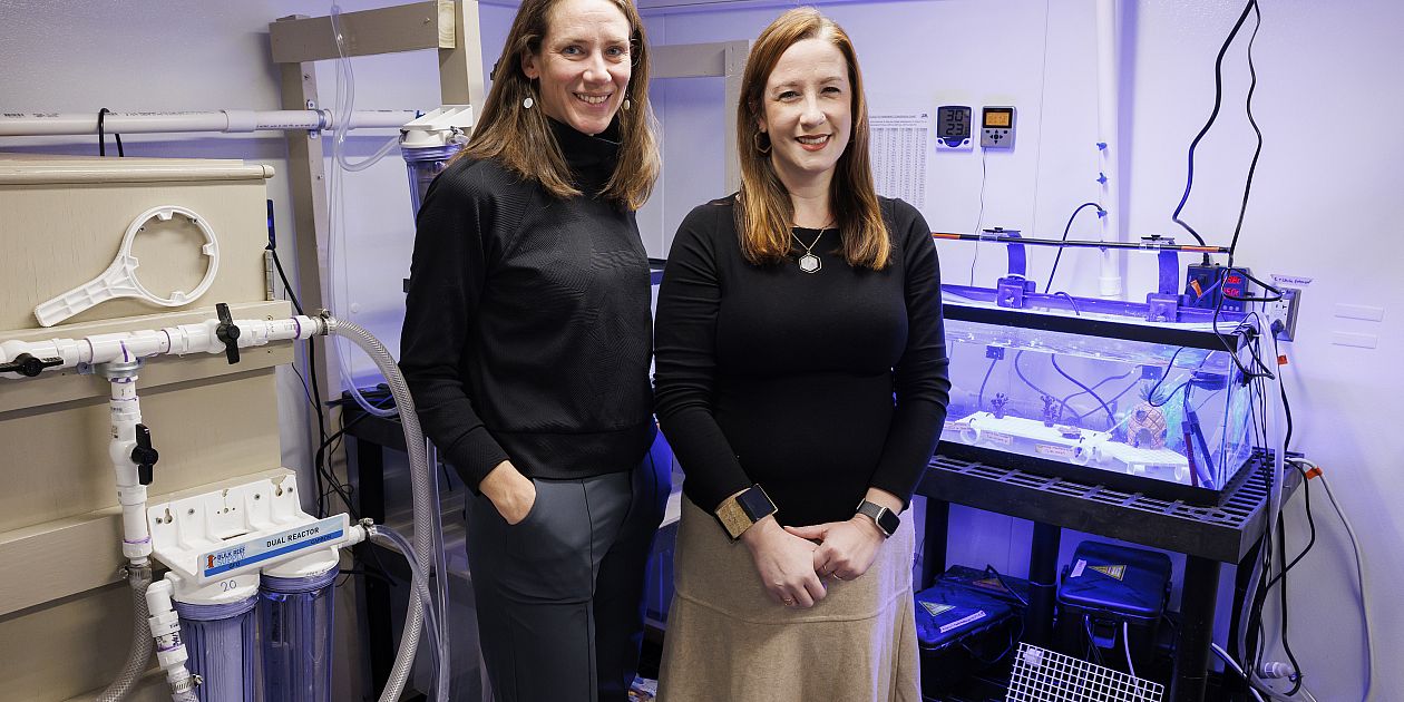
For nearly two decades, researcher Jesse Owens and his team at the University of Hawaiʻi at Mānoa John A. Burns School of Medicine (JABSOM) have been pursuing a bold…
A study published in the Lancet Public Health looks at oral nicotine pouch use in Great Britain.
Prof…

Introduction
An overview of the circadian rhythm
Why nighttime eating matters
Best foods to eat at night
Foods to limit or avoid in the evening
Effects on cardiometabolic health
Practical guidelines
References
Further reading
Late-night meals can…

According to the US Centers for Disease Control and Prevention (CDC) and other sources, there are approximately 1.2 million people living with HIV in the United States, and another 1.2 to 2.2 million who are at…
You don’t have permission to access “http://www.hhs.gov/press-room/cdc-adopts-individual-based-decision-making-for-hepatitis-b-immunization-for-infants-born-to-women-who-test-negative-for-the-hepatitis-b-virus.html” on this…

Vaccination in pregnancy (VIP) keeps parents and babies safer by preventing infection-related morbidity and mortality [-]. Despite these benefits, vaccine uptake in pregnancy remains low in Canada and globally, particularly after the…