15 Décembre 2025
The International Agency for Research on Cancer (IARC) was represented at “From Risk to Action: Preventing Liver Health Crises through NCD Literacy and Care”, a policy event on liver…
15 Décembre 2025
The International Agency for Research on Cancer (IARC) was represented at “From Risk to Action: Preventing Liver Health Crises through NCD Literacy and Care”, a policy event on liver…
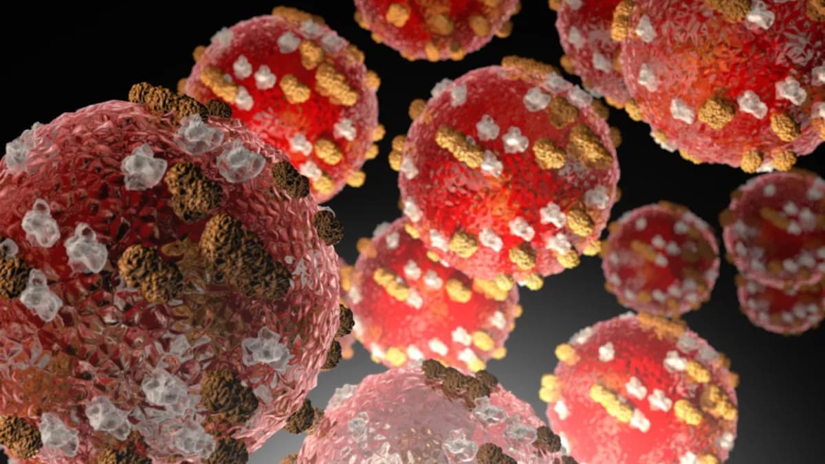
Liver cancer, especially hepatocellular carcinoma(HCC), is the fifth most common cancer worldwide and the third leading cause of cancer mortality.1 Asia is a major region for liver cancer, and Asia reported 72.5% of the world’s…

In the future, medications derived from cannabis may play a role in treating ovarian cancer. A research team examining two natural compounds found in cannabis discovered that both produced notable anti-cancer activity when tested on ovarian…
Chronic obstructive pulmonary disease (COPD), characterized by irreversible and progressive airflow limitation (AFL) due to chronic airway inflammation, and obstructive sleep apnea (OSA), defined by recurrent apnea and hypopnea…
Colorectal cancer (CRC) is the third most commonly diagnosed malignancy worldwide, accounting for approximately 10% of all new cancer cases and 9.4% of cancer-related deaths annually, making it the second leading cause of cancer…
Urinary incontinence (UI) is any involuntary leakage of urine,1 with a higher prevalence observed among older women.2 Globally, the prevalence of UI in older women ranged from 22% to 80%,3 with more than 25% of Chinese women aged…

Backed by major public investment, Ribeirão Preto’s Nutera Center launches a groundbreaking clinical program poised to cut treatment costs, expand access, and bring cutting-edge cancer care to the country’s public health system
1Department of Pathology, Cancer Hospital of China Medical University, Liaoning Cancer Hospital and Institute, Shenyang, 110000 People’s Republic of China; 2Department of Pathology, The 7th People’s Hospital of Zhengzhou, Zhengzhou…