Scientists have developed platelet-inspired nanoparticles that deliver anti-inflammatory drugs directly to brain-computer interface implants, doubling their effectiveness.

Scientists have found a way to improve the performance of…
Scientists have developed platelet-inspired nanoparticles that deliver anti-inflammatory drugs directly to brain-computer interface implants, doubling their effectiveness.
Scientists have found a way to improve the performance of…
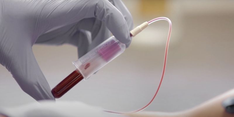
They’re approved by the FDA and Medicare and inching closer to widespread adoption nationally. But how good are they at detecting colorectal cancer?
Over the last several decades, a new mystery has emerged surrounding the rates of colorectal…

Updated on: Dec 11, 2025 06:17 pm IST
Granular parakeratosis (GP) is a relatively rare, benign disorder of keratinization. It typically presents clinically as scaly erythema or brownish hyperkeratotic papules and plaques, predominantly occurring in flexural or…
MOSCOW, Dec. 11 (Xinhua) — Russia’s first mRNA vaccine developed for melanoma therapy could be cleared for human use as early as 2026.
The Gamaleya National Research Center for Epidemiology and Microbiology has already produced several…
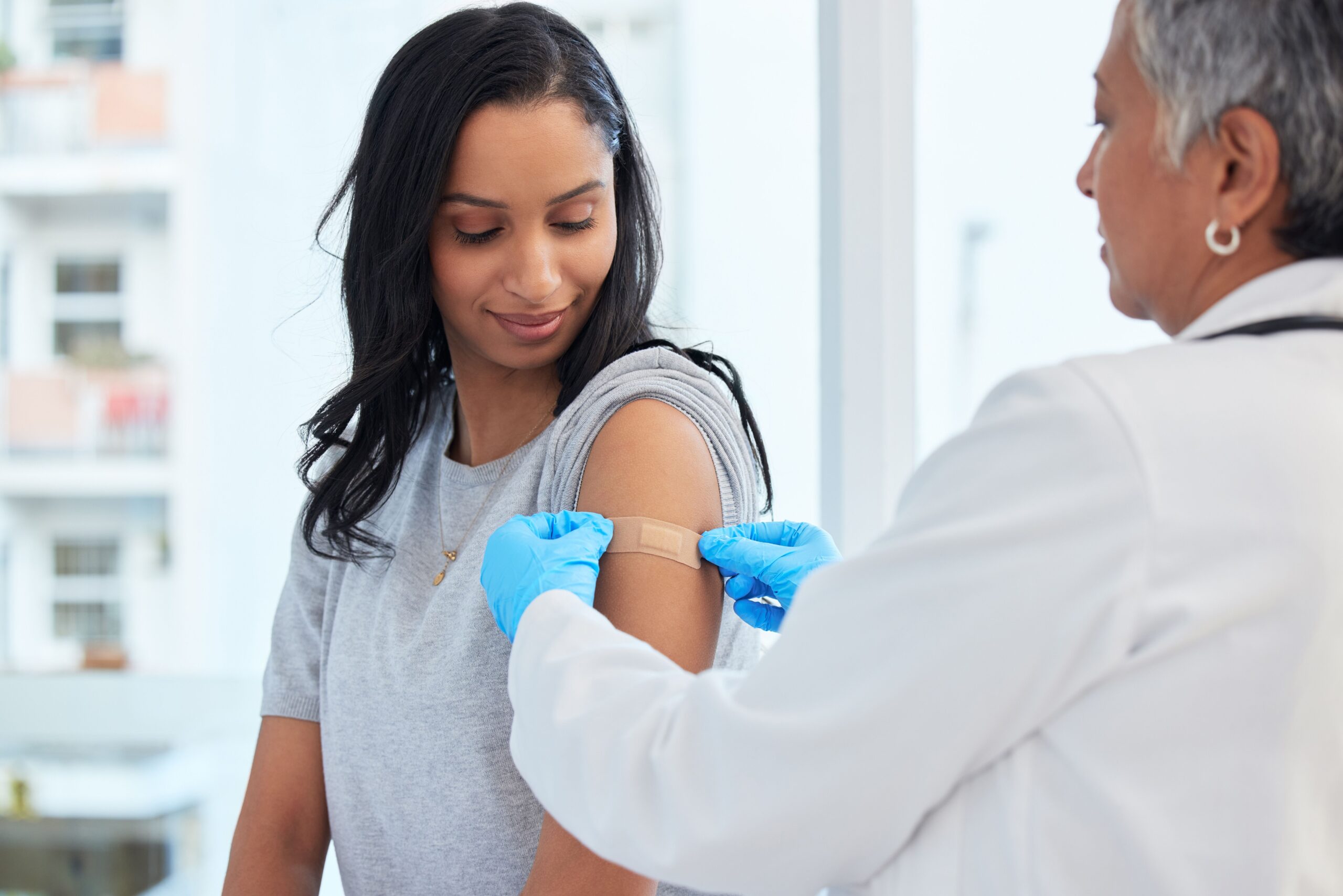
A phase 2, randomized, placebo-controlled study evaluating the investigational maternal vaccine GBS-AlpN found that the vaccine was immunogenic in pregnant individuals and resulted in high concentrations of vaccine-specific antibodies in infants…

Research suggests a surprising link between blood type and stroke risk, with people carrying one specific group A blood type facing a higher likelihood of stroke before age 60.
This discovery, published in a 2022 paper, deepens our…