Jen SmithSouth West health correspondent
 BBC
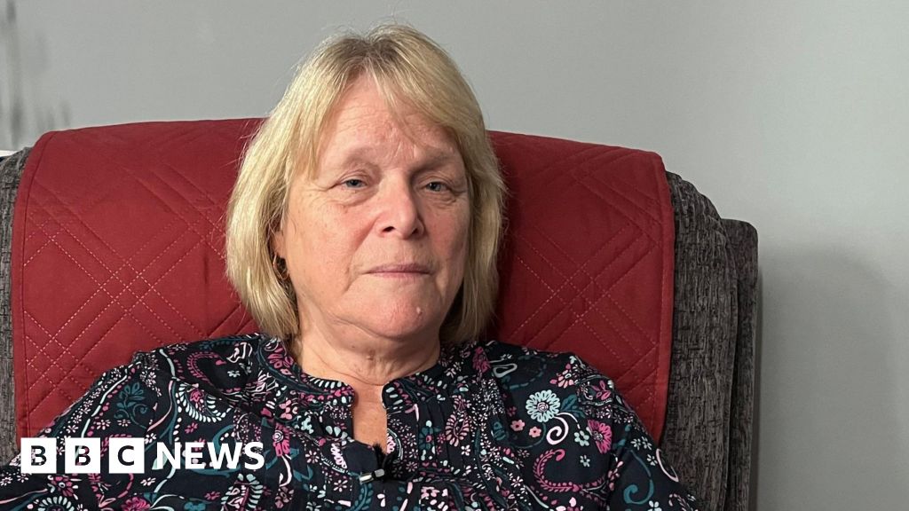
BBCA woman has said the weight loss drug Mounjaro has left her in chronic pain, suffering depression and with “no quality…
Jen SmithSouth West health correspondent
BBCA woman has said the weight loss drug Mounjaro has left her in chronic pain, suffering depression and with “no quality…

Antidepressants don’t have to be taken forever, a new analysis suggests.
Every year, a growing number of people across Europe take antidepressants to help treat symptoms related to depression and anxiety. While current guidelines recommend…
Cannabis use is rising among older adults, fueled by expanded legalization and people increasingly turning to cannabinoid-containing products to manage pain, sleep problems and…

The Guardian’s science editor, Ian Sample, sits down with co-host Madeleine Finlay to discuss three eye-catching stories from the week, including a study investigating the link between social media use in children and rising rates of ADHD…

Cancer drug resistance remains one of the biggest challenges in cancer treatment, and doctors urgently need better ways to prevent it. Yet scientists still do not fully understand the molecular processes that allow tumors to escape and return…
Cancer drug resistance remains one of the biggest challenges in cancer treatment, and doctors urgently need better ways to prevent it. Yet scientists still do not fully understand the molecular processes that allow tumors to escape and return…
1Emergency Trauma Center, The First Affiliated Hospital of Xinjiang Medical University, Urumqi, People’s Republic of China; 2Surgery for Hepatic and Biliary Echinococcosi, The First Affiliated Hospital of Xinjiang Medical University, Urumqi,…

Researchers at Georgetown University Medical Center have identified a way the brain’s learning system can shift depending on the activity of a particular protein. Their work shows that the ability to connect cues with rewarding outcomes can be…