Many of us are still in the reflective stage after
Category: 6. Health
-
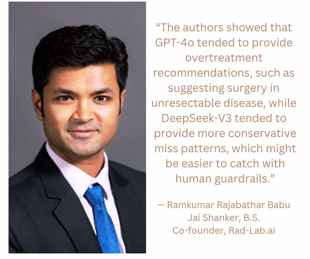
Clinical Applications of LLMs in Radiology: Key Takeaways from RSNA 2025
RSNA 2025 as we attempt to summarize all the fascinating AI-focused sessions, panels, and hallway discussions. Throughout the seven imaging informatics sessions this year, a clear message became… -

Tailoring Low-Protein Diets to Improve CKD Health Outcomes
Protein restriction has been used as a strategy for delaying disease progression in patients with
chronic kidney disease (CKD) for more than 140 years, said the authors of areview article covering research on a low-protein diet in animal models…Continue Reading
-

Gene Therapy Shows Promise Against T-ALL
Original story from the University College London (UCL; UK).
A groundbreaking new treatment using genome-edited immune cells, developed by scientists at UCL and Great Ormond Street Hospital (GOSH; London UK), has shown promising results in…
Continue Reading
-

Taken correctly, this mineral may shorten your cold – The Washington Post
- Taken correctly, this mineral may shorten your cold The Washington Post
- This popular supplement could shorten the duration of your cold The Independent
- What Happens to Your Cold Symptoms When You Take Vitamin C and Zinc Together Health: Trusted…
Continue Reading
-

Gray hair may have evolved as a protection against cancer, study hints
Graying hair could be a sign that the body is effectively protecting itself from cancer, a new study suggests.
Cancer-causing triggers, such as ultraviolet (UV) light or certain chemicals, activate a natural defensive pathway that leads to…
Continue Reading
-
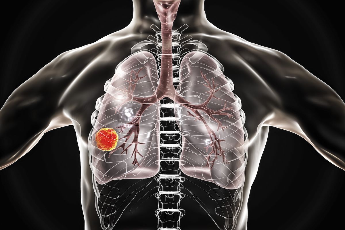
Ivonescimab/Chemo Improves Quality of Life in Frontline Squamous NSCLC
Updated results from the phase 3 HARMONi-6 trial (NCT05840016) demonstrated that patient-reported quality of life outcomes were improved with ivonescimab plus chemotherapy compared with tislelizumab-jsgr (Tevimbra) plus chemotherapy in the…
Continue Reading
-
Astrocyte Diversity Across Space and Time Mapped
When it comes to brain function, neurons get a lot of the glory. But healthy brains depend on the cooperation of many kinds of cells. The most abundant of the brain’s non-neuronal cells are astrocytes, star-shaped cells with a lot of…
Continue Reading
-

Australian researchers pinpoint specific genetic changes linked to severe AMD
Australian researchers have for the first time pinpointed specific genetic changes that increase the risk of severe, sight-threatening forms of age-related macular degeneration (AMD).
A new study, published today in Nature…
Continue Reading
-

Healthcare use patterns for high volume musculoskeletal shoulder disor
Introduction
Shoulder conditions and disorders are commonly experienced musculoskeletal pain conditions and have a prevalence (20.9%) comparable to back (26.9%) and neck pain (20.6%).1 For example, in a 15-year population-based study in the…
Continue Reading
-
UKHSA Detects New Recombinant Mpox Strain in England – Medscape
- UKHSA Detects New Recombinant Mpox Strain in England Medscape
- Inter-Clade Recombinant Mpox Virus Detected in England in a Traveller Recently Returned from Asia Virological
- People have been warned to stay alert facebook.com
- New recombinant…
Continue Reading