- Translating the Impact of the IMvigor011 Trial Readout: Reshaping Oncology Care Inside Precision Medicine
- Adjuvant Atezolizumab Generates DFS, OS Benefit Regardless of Tumor Size, Nodal Status, and Prior NAC in ctDNA+ MIBC OncLive
- Future…
Category: 6. Health
-
Translating the Impact of the IMvigor011 Trial Readout: Reshaping Oncology Care – Inside Precision Medicine
-

Study examines gestational diabetes in American Samoan women
Despite the high burden of disease, the underlying causes of GDM in Pacific Islanders remain poorly understood. GROW aims to change that. With investigators at the University of Pittsburgh, Emory University, and the Obesity, Lifestyle and Genetic…
Continue Reading
-
CONSUMING TREE NUTS AS SNACKS REDUCES FOOD CRAVINGS AND IMPROVES DIET QUALITY IN MILLENNIALS AT RISK FOR METABOLIC SYNDROME
New Findings Published in Nutrients in Partnership with The International Tree Nut Council Nutrition Research & Education Foundation
DAVIS, Calif., Dec. 9, 2025 /PRNewswire/ –…
Continue Reading
-
Dear Doctor: Can contrast dye used during MRIs cause symptoms of hyperthyroidism?
DEAR DR. ROACH: I am a male who is 58 years of age. I recently had an MRI of my cervical spine, and just this month, I had a CT scan of my whole abdomen. Since then, I have had symptoms of dry eyes, weight loss and insomnia. I wondered if the…
Continue Reading
-

Avant Technologies and Austrianova Advancing α-Klotho Cell Therapy as Mayo Clinic Study Links Low α-Klotho Levels to Poor Cardiovascular Survival
LAS VEGAS, Dec. 9, 2025 /PRNewswire/ — Avant Technologies, Inc. (OTCQB: AVAI) (“Avant” or the “Company”), an emerging biotechnology company focused on developing…
Continue Reading
-
Access to Vaccines – GPEI
The routine immunisation component is calculated using a standardised formula:
- Population × Scheduled Doses × Coverage × Wastage
The SIA component captures nOPV2 and bOPV requirements for outbreak response, endemic SIAs (in Afghanistan and…
Continue Reading
-

High BMI and poor physical fitness in adolescence linked to severe bacterial infections in adulthood
High BMI and poor physical fitness during later adolescence increase the risk of both contracting and dying from sepsis and other severe bacterial infections in adulthood, according to a study from the University of Gothenburg.
The…
Continue Reading
-
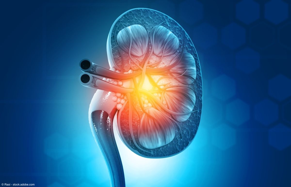
Neoadjuvant SAbR shows promising outcomes in RCC with tumor thrombus
In total, the prospective phase 2 study included 23 patients with newly diagnosed RCC. Patients received neoadjuvant SAbR as 40 Gy over 5 fractions or 36 Gy over 3 fractions, followed by surgery. The median follow-up was 35.2 months.
Data showed…
Continue Reading
-

Maternal Sleep Loss Linked to Offspring Ovarian Damage
A NEW mouse study suggests that severe sleep deprivation during early pregnancy could have lasting effects on daughters’ fertility by depleting their ovarian germ cell reserve through ferroptosis, an iron-dependent form of cell…
Continue Reading
-

Online reference library provides a more accurate picture of drug exposure
Doctors and researchers try to understand what medications a person has taken by asking patients directly or by looking at medical records. But this information is often incomplete. People may forget what they took, use…
Continue Reading