A recipient of a kidney transplant presented a medical mystery when he died from rabies in January 2025 only weeks after his surgery in an Ohio hospital, despite having had no documented contact with the disease.
A close investigation by the…
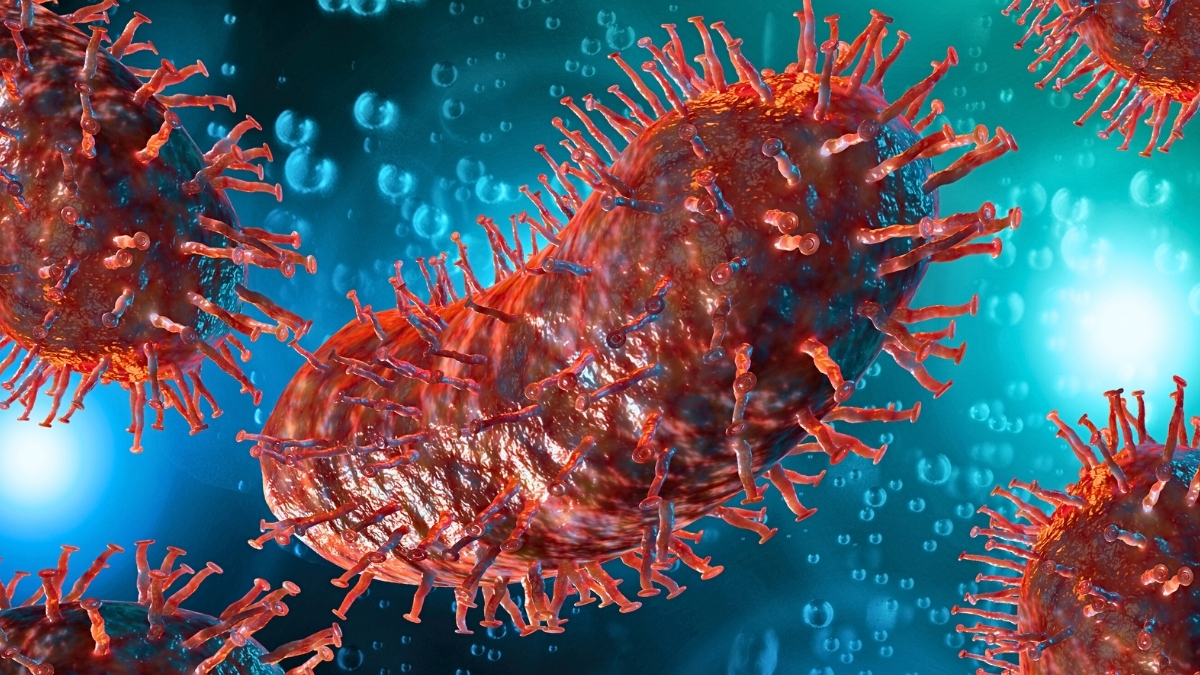
A recipient of a kidney transplant presented a medical mystery when he died from rabies in January 2025 only weeks after his surgery in an Ohio hospital, despite having had no documented contact with the disease.
A close investigation by the…
MELBOURNE, Dec. 8 (Xinhua) — Scientists in Australia have produced a first-of-its-kind catalog of pediatric cancers, offering fresh clues for developing targeted immunotherapies for young patients.
The Children’s Cancer Model Atlas (CCMA),…

Cold plunges have become a go-to ritual in modern wellness routines, praised for everything from boosting immunity to speeding up recovery. But here’s what often gets overlooked: most of the science behind these icy benefits is based on…
A study jointly carried out by the Infectious Hazard Management (IHM) Unit at WHO SEARO and the Institute of Development Studies (IDS), University of Sussex, introduces a practical framework and reflection tool to help national programme managers…

For more than a century, tuberculosis (TB), caused by the bacteria Mycobacterium tuberculosis, has remained a serious global health problem |Image used for representational purpose only
…

In a new leap for neurobiology and bioelectronics, Northwestern University scientists have developed a wireless device that uses light to send information directly to the brain – bypassing the body’s natural sensory pathways.
The soft,…
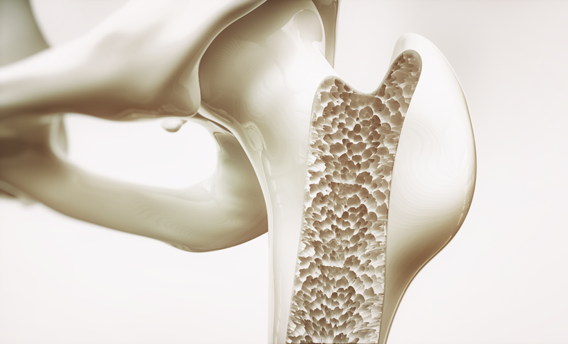
A NEW pilot trial suggests that adding low-frequency electrical stimulation to exercise may enhance local bone outcomes in premenopausal women at risk of osteoporosis.
The need for accessible…

Graphnet Health has been selected by Jersey’s Family Nursing & Home Care to provide the island’s first large-scale remote monitoring service.
The monitoring service is a landmark project funded through the Impact Jersey CareTech…
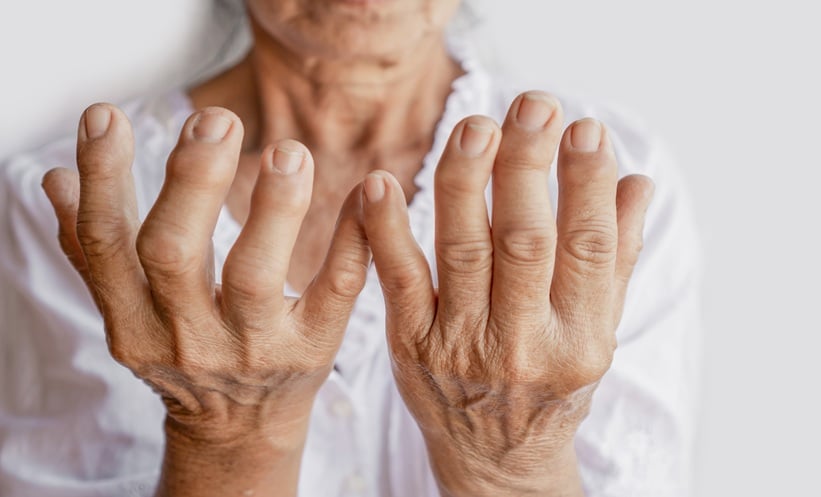
ACCELERATED biological aging may be a missing link between hyperuricemia, rising serum uric acid, gout, and diet-driven prevention.
In this large prospective analysis of 412,493 UK Biobank participants, investigators examined how biological…
