Researchers have discovered metabolites that impact liver metabolism and insulin sensitivity. They travel from the intestine to the liver and then to the heart, from where they are spread to the rest of the body.
The publication in Cell…

Researchers have discovered metabolites that impact liver metabolism and insulin sensitivity. They travel from the intestine to the liver and then to the heart, from where they are spread to the rest of the body.
The publication in Cell…

“You have weak digestion,” the reflexology therapist declared as she scraped a wooden massage stick up and down the arch in my right foot. I winced while nodding in agreement, half hoping she’d accept my admission as a peace offering and…
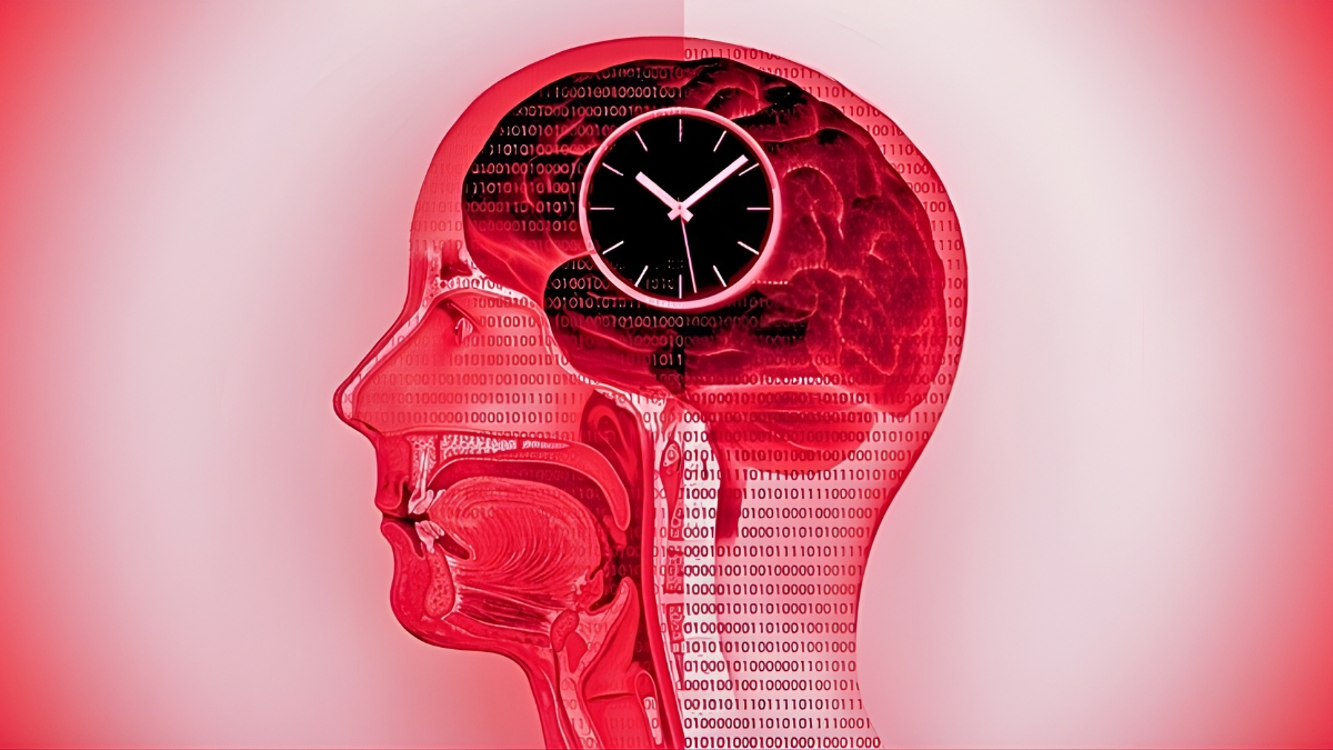
Scientists looking to tackle our ongoing obesity crisis have made an important discovery: Intermittent calorie restriction leads to significant changes both in the gut and the brain, which may open up new options for maintaining a healthy…

Scientists looking to tackle our ongoing obesity crisis have made an important discovery: Intermittent calorie restriction leads to significant changes both in the gut and the brain, which may open up new options for maintaining a healthy weight.
Continue Reading


The Hong Kong Centre for Health Protection (CHP) of the Department of Health reported an additional locally acquired chikungunya fever (CF) cases, bring the total to 10.
The case involves a 67-year-old female living in Kwai Tsing District. A…

Prof Geraldine Moses was speaking to a nurse who told her something concerning: patients with kidney failure were taking “iron supplements” that contained almost no iron.
Patients on kidney dialysis often need iron supplements because the…
By late life, about two thirds of people in Western countries develop an intestinal condition called Diverticular disease. It is rarely noticed, yet it can cause great harm.
Recent national surveys from the United Kingdom show that most adults…

FILE – A new study by UCLA Health found that people in emotionally supportive marriages have lower BMIs, healthier guts and higher levels of the “love hormone.”(Photo by OMAR KARIM/Middle East Images/AFP via Getty Images)
Emotionally…
