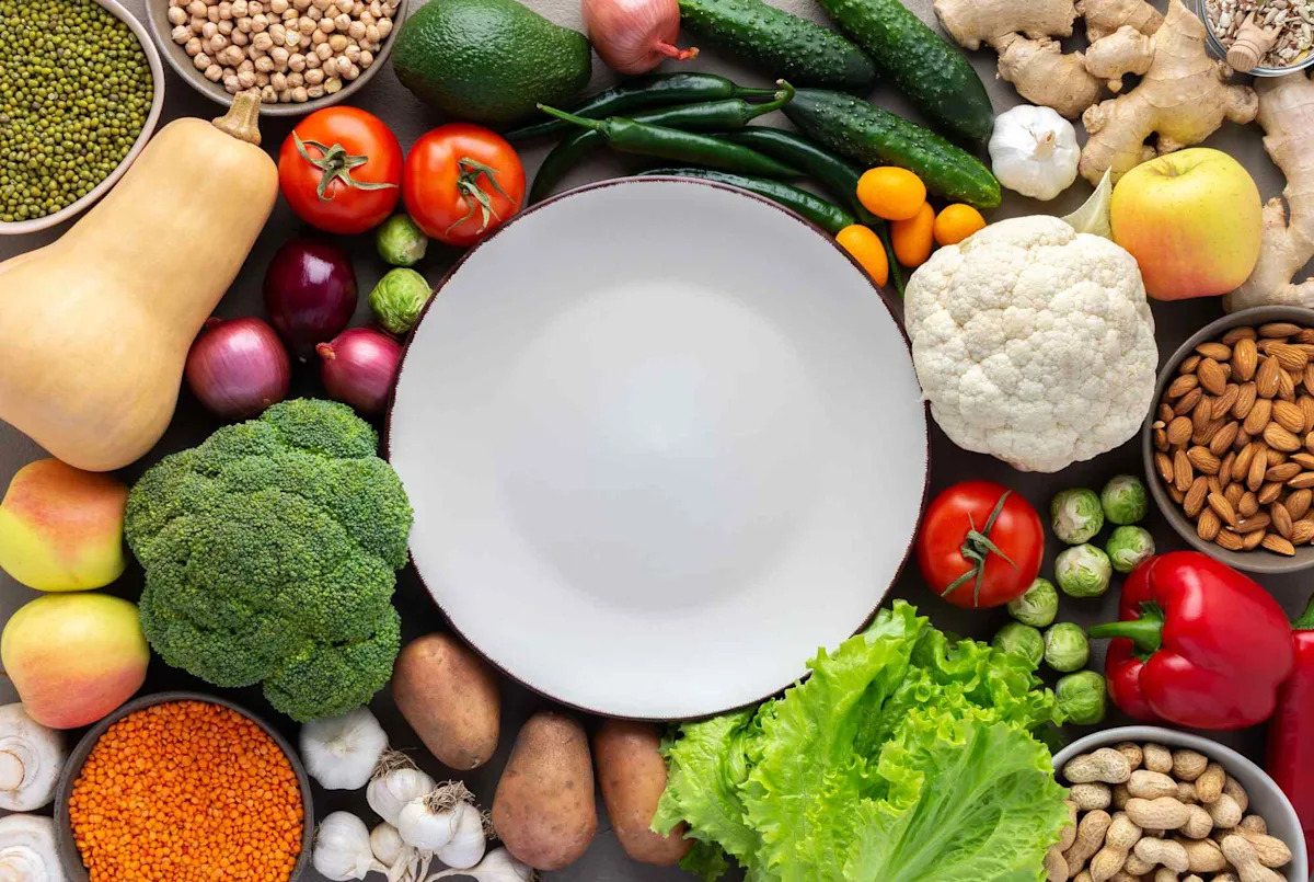
Category: 8. Health

  • They Thought It Was Heroin, But a Super Opioid 1000x Stronger Than Morphine Was Hiding Inside – SciTechDaily

    1. They Thought It Was Heroin, But a Super Opioid 1000x Stronger Than Morphine Was Hiding Inside  SciTechDaily
    2. Frankenstein Opioids: Drug Stronger Than Fentanyl Seized In All 50 States  Patch
    3. A New Drug More Potent Than Heroin and Fentanyl is Spreading Globally  Kursiv Media
    4. Highly potent opioid Nitazenes linked to growing number of overdose deaths  WBIW
    5. Nitazenes: A Hidden Threat Stronger Than Fentanyl  Conexiant

    Continue Reading

  • Massive Study Links 8 Genetic Signals to Chronic Fatigue Syndrome : ScienceAlert

    Massive Study Links 8 Genetic Signals to Chronic Fatigue Syndrome : ScienceAlert

    Chronic fatigue syndrome or myalgic encephalomyelitis (ME/CFS) is a debilitating and long-neglected disease that experts typically dismissed as psychosomatic for decades.

    To this day, some physicians still believe the illness is all in a patient’s head, but the largest genetic analysis of its kind suggests there are real biological origins.

    A UK research project, called DecodeME, has investigated the genome-wide associations and uncovered eight possible signals associated with ME/CFS.

    The preprint findings, which have not yet been published or peer-reviewed, suggest that an individual’s genes at least partly contribute to their chances of developing ME/CFS.

    Related: Long COVID Fatigue Shows Up as Distinct Changes in Brain Scans

    One of the eight genetic variants “nicely overlays” a signal that was previously linked to chronic pain, a common symptom of ME/CFS explains the project leader of DecodeME, bioinformaticist Chris Ponting from the University of Edinburgh.

    In addition, three of the eight signals are known to act as first responders to viral or bacterial infections. The findings could help explain why ME/CFS patients often report an infection before their first symptoms, and why numbers have surged since the pandemic.

    “DecodeME’s results, grounded in the principles of statistical genetics, now place ME/CFS research on a firm biological foundation,” concludes the DecodeME team of more than 50 researchers. This “should help to reduce the stigma of the illness,” they add.

    frameborder=”0″ allow=”accelerometer; autoplay; clipboard-write; encrypted-media; gyroscope; picture-in-picture; web-share” referrerpolicy=”strict-origin-when-cross-origin” allowfullscreen>

    Neuropsychiatrist Alan Carson from the University of Edinburgh, who was not involved in the study, says it is “by someway the largest study ever conducted on genetics of CFS/ME.”

    DecodeME’s analysis brought together more than 16,000 patients, most of whom are female and of European descent. The criteria for diagnosis was stringent so that only the most clear-cut cases were included.

    Patients must have had an official diagnosis of ME/CFS as well as a key symptom, called post-exertional malaise, which essentially translates to undue fatigue following exercise or other energy-consuming activities, including concentrating or socializing.

    The findings revealed over a dozen genetic signals linked to ME/CFS, but only eight could be replicated in a second dataset, including more than 13,000 cases.

    A third dataset of more than 14,000 cases failed to reproduce the results. This may have been due to differences in how ME/CFS was defined and diagnosed.

    While these eight genome-wide associations are not exclusive to those with ME/CFS, the findings suggest that they are more likely to occur in those with the disease.

    Many of the eight gene signals were expressed in brain tissue, including the ones associated with pain and the immune system.

    “Drugs targeting these genes’ proteins might help protect against the consequences of microbial infection and therefore could reduce the risk of acquiring ME/CFS,” the authors suggest.

    The signals are modest, and we don’t yet know what they mean. While there are limitations to the research, University of Hertfordshire geneticist Alena Pance, who was also not involved in the study, says the study is a “great advance towards understanding the illness better.”

    But while Carson agrees the research is important, he sees a long road ahead. Carson points out that in disorders like depression, finding a few associated genes has not advanced our understanding of the disease or improved treatments just yet.

    Still, important strides are being made with the help of patients. Ponting and his team argue that their recent findings help explain the heritable component of ME/CFS, improve the likelihood of finding effective drugs, and place the disease on more equal terms with other common genetic conditions.

    “We’ve gone from knowing almost nothing about the causes of ME to having specific genetic information to delve much deeper into,” says study contributor Sonya Chowdhury, the CEO of UK health charity, Action for ME.

    “For decades people with ME have asked to be heard, and now science is catching up.”

    The research is available via preprint here.

    Continue Reading

  • Lung Cancer Predictive Models | RSNA

    Predictive models for lung cancer often fall short when applied beyond the clinical settings in which they were developed, especially when evaluating biopsied lung nodules.

    A recent study in Radiology: Artificial Intelligence highlights this challenge and offers guidance for improving models’ generalizability across institutions and clinical settings with strategies such as image harmonization and fine-tuning models on local patient populations.

    More than 1.5 million Americans have at least one pulmonary nodule detected either incidentally at routine chest CT or during lung cancer screening every year. Nodule biopsy carries risks, costs and anxiety for patients. With 95% of indeterminate pulmonary nodules found to be benign, clinical guidelines recommend risk-stratifying nodules before resorting to invasive percutaneous or surgical interventions.

    “We want to diagnose these pulmonary nodules earlier and noninvasively, and we want to avoid performing a biopsy on benign nodules,” said study lead author Thomas Z. Li, PhD, from the Medical-image Analysis and Statistical Interpretation (MASI) Lab at Vanderbilt University in Nashville, TN. “Better noninvasive diagnostic tools can help us do that.”

    Statistical models for predicting lung cancer have the potential to improve risk stratification, aiding in earlier diagnosis of malignancy as well as reducing the risk of morbidity, costs and unnecessary anxiety associated with the workup of benign disease. Several models have been validated, but a systematic analysis of their performance is lacking.

    To learn more, Dr. Li and colleagues evaluated eight validated predictive models developed to stratify pulmonary nodules. The models consisted of clinical prediction models, cross-sectional or longitudinal AI models, and multimodal approaches. The researchers evaluated the models on nine patient cohorts in three clinical settings: nodules detected during screening, incidentally detected nodules and pulmonary nodules deemed suspicious enough to warrant a biopsy.

    “We wanted to know, in these three clinical settings, how do the models that have been developed so far perform?” Dr. Li said.

    Analysis revealed that the eight lung cancer prediction models failed to generalize well across clinical settings and sites outside of their training distributions.

    The findings show that a single external validation set is not enough to guarantee generalization performance, Dr. Li noted.

    “You’re training the model on one group, which is a healthy screening population, and then you’re trying to apply it into a different group, and what we see is that it doesn’t work,” he said. “We need the model to be evaluated across multiple different institutions, and we need it to be evaluated in different clinical settings.”

    Continue Reading

  • Shanelle’s story: The power of representation in clinical trials

    Shanelle’s story: The power of representation in clinical trials

    Lupus is a chronic autoimmune disease which disproportionately impacts women of childbearing age, particularly women of color.1 It is two to three times more prevalent among African American, Hispanic/Latina, Asian American, Native American, Alaska Native, Native Hawaiian and other Pacific Islander women than among White women.2 In the U.S, about 90% of people living with lupus are women, and most people experience initial symptoms between ages 15 and 44.1 Despite its prevalence, treatment options for lupus have long been limited.

    The epidemiology of those living with lupus underscores why representative participation in clinical trials is so vital: New therapies should include the patient populations that are affected by the disease to ensure the therapies are effective. Shanelle also recognizes it’s not just about clinical trial enrollment, but about the entire experience.  

    “One of the challenges in the clinical trial process is the lack of diversity in staffing and accessibility within the communities that need it most. For a lot of people, it means something to have somebody that culturally relates to them,” Shanelle explained. Today, she is a vocal advocate who works to raise awareness of lupus and its effects, particularly on women of color. 

    Continue Reading

  • Research links chocolate consumption to improved blood circulation – Samaa TV

    1. Research links chocolate consumption to improved blood circulation  Samaa TV
    2. Consumption of Flavan-3-ol-Rich Foods may help Lower Blood Pressure: Study  Medical Dialogues
    3. Five everyday foods that could help lower blood pressure – including dark chocolate  Manchester Evening News
    4. Skincare benefits of dark chocolates, why it should be a part of your diet to keep skin healthy  Moneycontrol
    5. Can Dark Chocolate Boost Good Cholesterol? Here’s What Science Says  TheHealthSite

    Continue Reading

  • Genetic Testing May Reduce Chemotherapy Side Effects for Patients With GI Cancers

    Genetic Testing May Reduce Chemotherapy Side Effects for Patients With GI Cancers

    For patients with gastrointestinal (GI) cancers, chemotherapy can sometimes cause severe, even life-threatening side effects in those who carry certain genetic variants that may impact how their bodies process the drugs used to treat their disease. Testing for variants in two genes before starting chemotherapy may significantly improve patient safety by providing physicians with information to help tailor doses, according to new research from the Perelman School of Medicine at the University of Pennsylvania, published by Tuteja et al in JCO Precision Oncology. The study showed that dose adjustments based on preemptive genetic testing cut chemotherapy side effects in half, as compared with patients who have the genetic variants and received standard doses without prior testing.

    “For too long, the [United States] lagged behind Europe in adopting genetic testing for chemotherapy dosing, but our study shows it’s not only feasible but also critical for patient safety,” said the study’s lead author Sony Tuteja, PharmD, MS, Director of Pharmacogenomics in the Penn Medicine Center for Genomic Medine and Research Assistant Professor of Translational Medicine and Human Genetics. “With up to 1,300 deaths in the U.S. each year due to side effects from one of the most common forms of chemotherapy drugs, we’ve worked to make testing fast and actionable, getting results in about a week to help doctors make safer treatment decisions.”

    Nearly 290,000 Americans are diagnosed with GI cancers each year—including colorectal cancer, the third most common cancer diagnosis in the nation. Current chemotherapy protocols use standard dosing standards that do not account for genetic differences in how patients process these drugs.

    Genetic Variants Guide Safer Chemotherapy

    The study focused on variants in two genes: DPYD and UGT1A1. The DPYD gene produces an enzyme that helps the liver break down drugs like fluoropyrimidines, which are commonly used in GI cancer treatment. About 5% to 8% of people carry DPYD variants that hinder the body’s ability to process fluoropyrimidine, causing it to build up to harmful levels, which can lead to serious side effects like reduced blood cell production, mouth sores, or hand-foot syndrome. Similarly, the UGT1A1 gene affects how the body processes irinotecan, another key chemotherapy drug often used to treat GI cancers. Variants in UGT1A1 can lead to the body processing the drug too slowly, increasing the risk of severe diarrhea or low white blood cell counts. By identifying these variants, physicians can lower chemotherapy doses to prevent harmful side effects without compromising treatment effectiveness.

    The study enrolled 517 patients with GI cancer at three cancer care sites in the University of Pennsylvania Health System who were scheduled to begin treatment with fluoropyrimidine or irinotecan. A group of 288 received blood tests to check for DPYD and UGT1A1 variants.

    Among 16 patients who were found to have genetic variants and received tailored dose reductions based on test results, 38% experienced severe treatment-related adverse events. In comparison, 65% of 17 patients with the genetic variants from a biobank group who received standard doses without prior testing experienced the serious side effects. The tested group also saw a significantly lower need to change treatment dosage and frequency (38% vs 76%) and fewer treatment discontinuations (31% vs 47%).

    Disclosure: The research was funded in part by grants from the Penn Center for Precision Medicine and the National Institute of Health’s National Center for Advancing Translational Sciences. For full disclosures of the study authors, visit ascopubs.org.

    Continue Reading

  • Faryabi R, Kamali A, Raesi R, Hushmandi K, Tabatabaee SS, Askarpour H, Samakkhah SA, Daneshi S. Effects of COVID-19 pandemic on pulmonary tuberculosis (TB) occurrence in the South of Iran. Curr Respiratory Med Reviews. 2025;21(3):267–75.

    CAS 

    Google Scholar 

  • Organization WH. Global tuberculosis report 2021: supplementary material. World Health Organization; 2022.

    Google Scholar 

  • Lee SH. Tuberculosis infection and latent tuberculosis. Tuberc Respir Dis. 2016;79(4):201.

    Google Scholar 

  • Faryabi R, Raesi R, Taghdisi Heydarian SM, Daneshi S, Kermani B, Hushmandi K, Barfar E. Evaluation of the tuberculosis surveillance system in the Southern Region of Iran. Open Public Health J. 2024;17(1):e18749445275993.

    Google Scholar 

  • Talip BA, Sleator RD, Lowery CJ, Dooley JS, Snelling WJ. An update on global tuberculosis (TB). Infect Diseases: Res Treat. 2013;6:IDRT. S11263.

    Google Scholar 

  • Puyat JH, Brode SK, Shulha H, Romanowski K, Menzies D, Benedetti A, Duchen R, Huang A, Fang J, Macdonald L. Predicting risk of tuberculosis (TB) disease in people who migrate to a Low-TB incidence country: development and validation of a multivariable, dynamic risk-Prediction model using health administrative data. Clin Infect Dis. 2025;80(3):644–52.

    PubMed 

    Google Scholar 

  • Houben RM, Dodd PJ. The global burden of latent tuberculosis infection: a re-estimation using mathematical modelling. PLoS Med. 2016;13(10):e1002152.

    PubMed 
    PubMed Central 

    Google Scholar 

  • Gustafsson N, Leino-Kilpi H, Prga I, Suhonen R, Stolt M. Action–CA15208 RcC: Missed care from the patient’s perspective–a scoping review. Patient Prefer Adherence. 2020;14:383–400.

    PubMed 
    PubMed Central 

    Google Scholar 

  • Chakaya J, Petersen E, Nantanda R, Mungai BN, Migliori GB, Amanullah F, Lungu P, Ntoumi F, Kumarasamy N, Maeurer M. The WHO global tuberculosis 2021 Report–not so good news and turning the tide back to end TB. Int J Infect Dis. 2022;124:S26–9.

    PubMed 
    PubMed Central 

    Google Scholar 

  • Bagcchi S. WHO’s global tuberculosis report 2022. Lancet Microbe. 2023;4(1):e20.

    PubMed 

    Google Scholar 

  • Rastegar M, Nazar E, Nasehi M, Sharafi S, Fakoor V, Shakeri MT. Bayesian Estimation of the time-varying reproduction number for pulmonary tuberculosis in iran: a registry-based study from 2018 to 2022 using new smear-positive cases. Infect Disease Modelling. 2024;9(3):963–74.

    Google Scholar 

  • Song H-W, Tian J-H, Song H-P, Guo S-J, Lin Y-H, Pan J-S. Tracking multidrug resistant tuberculosis: a 30-year analysis of global, regional, and National trends. Front Public Health. 2024;12:1408316.

    PubMed 
    PubMed Central 

    Google Scholar 

  • Srivastava V, Verma A. Current challenges in the management of tuberculosis. J Young Pharmacists. 2024;16(2):145–54.

    CAS 

    Google Scholar 

  • Hassan AO. Social support and treatment outcomes among multi-drug-resistant tuberculosis and persons living with HIV in Nigeria. Walden University; 2025.

    Google Scholar 

  • Ehsani R, Sharafoddin M, Olyaeemanesh A, Delavari A, Vosoogh-Moghaddam A, Alimohamadi Y, Ghorbani S. Insights into health program implementation and evaluation in iran: A brief review of National healthcare programs in Iran. Iran J Public Health. 2024;53(11):2572.

    PubMed 
    PubMed Central 

    Google Scholar 

  • Nasiri MJ, Dabiri H, Darban-Sarokhalil D, Rezadehbashi M, Zamani S. Prevalence of drug-resistant tuberculosis in iran: systematic review and meta-analysis. Am J Infect Control. 2014;42(11):1212–8.

    PubMed 

    Google Scholar 

  • Ayoubi S, Farnia P, Farnia P, Ghanavi J, Velayati AA. Prevalence and Temporal trends of multidrug-resistant tuberculosis in Iran from 1981 to 2023: a systematic review and meta-analysis. Int J Mycobacteriology. 2024;13(3):320–30.

    Google Scholar 

  • Chowdhury K, Ahmad R, Sinha S, Dutta S, Haque M. Multidrug-resistant TB (MDR-TB) and extensively drug-resistant TB (XDR-TB) among children: where we stand now. Cureus. 2023;15(2):e35154.

    PubMed 
    PubMed Central 

    Google Scholar 

  • Koch A, Cox H, Mizrahi V. Drug-resistant tuberculosis: challenges and opportunities for diagnosis and treatment. Curr Opin Pharmacol. 2018;42:7–15.

    CAS 
    PubMed 
    PubMed Central 

    Google Scholar 

  • Dookie N, Ngema SL, Perumal R, Naicker N, Padayatchi N, Naidoo K. The changing paradigm of drug-resistant tuberculosis treatment: successes, pitfalls, and future perspectives. Clin Microbiol Rev. 2022;35(4):e00180–00119.

    PubMed 
    PubMed Central 

    Google Scholar 

  • Chen Z, Wang T, Du J, Sun L, Wang G, Ni R, An Y, Fan X, Li Y, Guo R. Decoding the WHO global tuberculosis report 2024: A critical analysis of global and Chinese key data. Zoonoses. 2025;5(1):999.

    CAS 

    Google Scholar 

  • Stephanie F, Saragih M, Tambunan USF. Recent progress and challenges for drug-resistant tuberculosis treatment. Pharmaceutics. 2021;13(5):592.

    CAS 
    PubMed 
    PubMed Central 

    Google Scholar 

  • Bialvaei AZ, Asgharzadeh M, Aghazadeh M, Nourazarian M, Kafil HS. Challenges of tuberculosis in Iran. Jundishapur J Microbiol. 2017;10(3):e37866.

    Google Scholar 

  • Madadi-Goli N, Ahmadi K, Meskini M, Vaziri F, Fateh A, Siadat SD. Tuberculosis prevention, diagnosis, and treatment financial profile during 2006–2021: part B. J Prev Diagn Treat Strategies Med. 2024;3(1):37–43.

    Google Scholar 

  • Moosazadeh M, Amiresmaili M. Challenges in case finding of tuberculosis control program in iran: A qualitative study. Bangladesh J Med Sci. 2018;17(3):462–9.

    Google Scholar 

  • Kazerooni P, Nejat M, Akbarpoor M, Sedaghat Z, Fararouei M. Underascertainment, underreporting, representativeness and timeliness of the Iranian communicable disease surveillance system for tuberculosis. Public Health. 2019;171:50–6.

    CAS 
    PubMed 

    Google Scholar 

  • Khademi F, Yousefi-Avarvand A, Derakhshan M, Vaez H, Sadeghi R. Middle East Mycobacterium tuberculosis antibiotic resistance: a systematic review and meta-analysis. Infect Epidemiol Microbiol. 2017;3(1):25–35.

    Google Scholar 

  • Abbasian S, Heidari H, Tadi DA, Kardan-Yamchi J, Taji A, Darbandi A, Asadollahi P, Maleki A, Kazemian H. Epidemiology of first-and second-line drugs-resistant pulmonary tuberculosis in iran: systematic review and meta-analysis. J Clin Tuberculosis Other Mycobact Dis. 2024;35:100430.

    CAS 

    Google Scholar 

  • Bahraminia F, Azimi T, Zangiabadian M, Nasiri MJ, Goudarzi M, Dadashi M, Fooladi AAI. Rifampicin-resistant tuberculosis in iran: A systematic review and meta-analysis. Iran J Basic Med Sci. 2021;24(6):720.

    PubMed 
    PubMed Central 

    Google Scholar 

  • Alibrahim A, Alqahtani H, Thirunavukkarasu A, Qazi I. Prevalence, patterns, and determinants of drug-resistant tuberculosis in Gulf Cooperation Council countries: an updated systematic review. PeerJ. 2024;12:e18628.

    PubMed 
    PubMed Central 

    Google Scholar 

  • Munir MA, Khan S, Rehman S, Ahmed D, Jabbar A. Tuberculosis among diabetes patients: a review of epidemiology, pathophysiology, clinical manifestations, and management. Chronicles Biomedical Sci. 2024;1(3):PID26–26.

    Google Scholar 

  • Page MJ, McKenzie JE, Bossuyt PM, Boutron I, Hoffmann TC, Mulrow CD, Shamseer L, Tetzlaff JM, Akl EA, Brennan SE. The PRISMA 2020 statement: an updated guideline for reporting systematic reviews. BMJ. 2021;372:n71.

    PubMed 
    PubMed Central 

    Google Scholar 

  • Schardt C, Adams MB, Owens T, Keitz S, Fontelo P. Utilization of the PICO framework to improve searching pubmed for clinical questions. BMC Med Inf Decis Mak. 2007;7:1–6.

    Google Scholar 

  • Sguanci M, Mancin S, Piredda M, De Marinis MG. Protocol for conducting a systematic review on diagnostic accuracy in clinical research. MethodsX. 2024;12:102569.

    PubMed 
    PubMed Central 

    Google Scholar 

  • Munn Z, Barker TH, Moola S, Tufanaru C, Stern C, McArthur A, Stephenson M, Aromataris E. Methodological quality of case series studies: an introduction to the JBI critical appraisal tool. JBI Evid Synthesis. 2020;18(10):2127–33.

    Google Scholar 

  • Sguanci M, Ferrara G, Palomares SM, Parozzi M, Godino L, Gazineo D, Anastasi G, Mancin S. Dysgeusia and chronic kidney disease: a scoping review. J Ren Nutr. 2024;34:374.

    CAS 
    PubMed 

    Google Scholar 

  • Howick J. The Oxford 2011 levels of evidence. http://www.cebm.net /index aspx? o = 5653 2011.

  • Mancin S, Sguanci M, Andreoli D, Soekeland F, Anastasi G, Piredda M, De Marinis MG. Systematic review of clinical practice guidelines and systematic reviews: a method for conducting comprehensive analysis. MethodsX. 2024;12:102532.

    PubMed 

    Google Scholar 

  • Mohajeri P, Sadri H, Farahani A, Norozi B, Atashi S. Frequency of mutations associated with rifampicin resistance in Mycobacterium tuberculosis strains isolated from patients in West of Iran. Microb Drug Resist. 2015;21(3):315–9.

    CAS 
    PubMed 

    Google Scholar 

  • Tabarsi P, Chitsaz E, Baghaei P, Shamaei M, Farnia P, Marjani M, Kazempour M, Amiri M, Mansouri D, Masjedi MR. Impact of extensively drug-resistant tuberculosis on treatment outcome of multidrug-resistant tuberculosis patients with standardized regimen: report from Iran. Microb Drug Resist. 2010;16(1):81–6.

    CAS 
    PubMed 

    Google Scholar 

  • Velayati AA, Masjedi MR, Farnia P, Tabarsi P, Ghanavi J, ZiaZarifi AH, Hoffner SE. Emergence of new forms of totally drug-resistant tuberculosis bacilli: super extensively drug-resistant tuberculosis or totally drug-resistant strains in Iran. Chest. 2009;136(2):420–5.

    PubMed 

    Google Scholar 

  • Mohajeri P, Moradi S, Atashi S, Farahani A. Mycobacterium tuberculosis Beijing genotype in Western iran: distribution and drug resistance. J Clin Diagn Research: JCDR. 2016;10(10):DC05.

    CAS 

    Google Scholar 

  • Doustdar F, Khosravi AD, Farnia P, Masjedi MR, Velayati AA. Molecular analysis of Isoniazid resistance in different genotypes of Mycobacterium tuberculosis isolates from Iran. Microb Drug Resist. 2008;14(4):273–9.

    CAS 
    PubMed 

    Google Scholar 

  • Lee H, Kim J, Kim J, Park Y-J. Review of the global burden of tuberculosis in 2023: insights from the WHO Global Tuberculosis Report 2024. 2025.

  • Tuberculosis. incidence in Iran. https://www.theglobaleconomy.com/Iran/Tuberculosis/?utm_source=chatgpt.com

  • Ebrahimzadeh A, Pagheh AS, Mousavi T, Fathi M, Moghaddam SGM. Serosal membrane tuberculosis in iran: A comprehensive review of evidences. J Clin Tuberculosis Other Mycobact Dis. 2023;31:100354.

    CAS 

    Google Scholar 

  • Yuanyuan L, Jingjing X, Shutao L, Dongdong S, Tian Z, Puxuan L, Yi P. 2024 WHO tuberculosis report: key data analysis for China and the global world. Electron J Emerg Infect Dis. 2025;9(6):92.

    Google Scholar 

  • Zumla A, George A, Sharma V, Herbert N. WHO’s 2013 global report on tuberculosis: successes, threats, and opportunities. Lancet. 2013;382(9907):1765–7.

    PubMed 

    Google Scholar 

  • Gizaw Z, Astale T, Kassie GM. What improves access to primary healthcare services in rural communities? A systematic review. BMC Prim Care. 2022;23(1):313.

    PubMed 
    PubMed Central 

    Google Scholar 

  • Douthit N, Kiv S, Dwolatzky T, Biswas S. Exposing some important barriers to health care access in the rural USA. Public Health. 2015;129(6):611–20.

    CAS 
    PubMed 

    Google Scholar 

  • Yu SW, Hill C, Ricks ML, Bennet J, Oriol NE. The scope and impact of mobile health clinics in the united states: a literature review. Int J Equity Health. 2017;16:1–12.

    Google Scholar 

  • Javanparast S, Windle A, Freeman T, Baum F. Community health worker programs to improve healthcare access and equity: are they only relevant to low-and middle-income countries? Int J Health Policy Manage. 2018;7(10):943.

    Google Scholar 

  • Nunemo MH, Gidebo KD, Woticha EW, Lemu YK. Integration challenges and opportunity of implementing non-communicable disease screening intervention with tuberculosis patient care: A mixed implementation study. Risk Manage Healthc Policy. 2023;16:2609–33.

    Google Scholar 

  • Arini M, Sugiyo D, Permana I. Challenges, opportunities, and potential roles of the private primary care providers in tuberculosis and diabetes mellitus collaborative care and control: a qualitative study. BMC Health Serv Res. 2022;22(1):215.

    PubMed 
    PubMed Central 

    Google Scholar 

  • Wagnew F, Gray D, Tsheten T, Kelly M, Clements AC, Alene KA. Effectiveness of nutritional support to improve treatment adherence in patients with tuberculosis: a systematic review. Nutr Rev. 2024;82(9):1216–25.

    PubMed 

    Google Scholar 

  • Saidi SS, Abdul Manaf R. Effectiveness of family support health education intervention to improve health-related quality of life among pulmonary tuberculosis patients in melaka, Malaysia. BMC Pulm Med. 2023;23(1):139.

    PubMed 
    PubMed Central 

    Google Scholar 

  • Palomino JC, Martin A. Drug resistance mechanisms in Mycobacterium tuberculosis. Antibiotics. 2014;3(3):317–40.

    PubMed 
    PubMed Central 

    Google Scholar 

  • Ramirez MV, Cowart KC, Campbell PJ, Morlock GP, Sikes D, Winchell JM, Posey JE. Rapid detection of Multidrug-Resistant Mycobacterium tube rculosis by use of Real-Time PCR and High-Resolution melt analysis. J Clin Microbiol. 2010;48(11):4003–9.

    CAS 
    PubMed 
    PubMed Central 

    Google Scholar 

  • Ong DC, Yam W-C, Siu GK, Lee AS. Rapid detection of rifampicin-and isoniazid-resistant Mycobacterium tuberculosis by high-resolution melting analysis. J Clin Microbiol. 2010;48(4):1047–54.

    CAS 
    PubMed 
    PubMed Central 

    Google Scholar 

  • Yin X, Zheng L, Liu Q, Lin L, Hu X, Hu Y, Wang Q. High-resolution melting curve analysis for rapid detection of Rifampin resistance in Mycobacterium tuberculosis: a meta-analysis. J Clin Microbiol. 2013;51(10):3294–9.

    CAS 
    PubMed 
    PubMed Central 

    Google Scholar 

  • Walker TM, Kohl TA, Omar SV, Hedge J, Elias CDO, Bradley P, Iqbal Z, Feuerriegel S, Niehaus KE, Wilson DJ. Whole-genome sequencing for prediction of Mycobacterium tuberculosis drug susceptibility and resistance: a retrospective cohort study. Lancet Infect Dis. 2015;15(10):1193–202.

    CAS 
    PubMed 
    PubMed Central 

    Google Scholar 

  • Sheikh BA, Bhat BA, Mehraj U, Mir W, Hamadani S, Mir MA. Development of new therapeutics to Meet the current challenge of drug resistant tuberculosis. Curr Pharm Biotechnol. 2021;22(4):480–500.

    CAS 
    PubMed 

    Google Scholar 

  • Terreni M, Taccani M, Pregnolato M. New antibiotics for multidrug-resistant bacterial strains: latest research developments and future perspectives. Molecules. 2021;26(9):2671.

    CAS 
    PubMed 
    PubMed Central 

    Google Scholar 

Continue Reading

  • Study Reveals Cholesterol’s Role in Fibrotic Progression of Metabolic Liver Disease

    Study Reveals Cholesterol’s Role in Fibrotic Progression of Metabolic Liver Disease

    Gerald I. Shulman, MD, PhD

    Cholesterol plays a vital role in the body, providing structural support to cells and serving as a building block for hormone synthesis. However, when cholesterol accumulates or is improperly distributed, it can contribute to the development and progression of disease. In a new study, published in Proceedings of the National Academy of Sciences, Yale School of Medicine (YSM) researchers showed that excess cholesterol stored in the liver can directly drive fibrosis in the context of metabolic disease.

    Led by Gerald I. Shulman, MD, PhD, the study aimed to identify key molecular triggers of metabolic dysfunction–associated steatohepatitis (MASH), a progressive liver disease marked by fat accumulation, inflammation, and fibrosis. According to Shulman, George R. Cowgill Professor of Medicine (Endocrinology) and professor of cellular and molecular physiology at YSM, understanding the drivers of fibrosis is critical to improving outcomes for patients with MASH.

    “Once fibrosis develops, it becomes very difficult to reverse and may ultimately progress to end-stage liver disease,” he says.

    Interestingly, says Shulman, it wasn’t the total amount of cholesterol in the liver that mattered most, but rather where it was stored. Specifically, cholesterol accumulated within liver fat droplets emerged as a key driver of liver inflammation and fibrosis.

    “It’s not just how much cholesterol is present, it’s about where it ends up,” says Shulman. “In this case, it was the cholesterol in the lipid droplets that triggers the damage. When it comes to lipids and liver disease, it’s all about location.”

    The researchers first identified this link using preclinical models and then validated their findings in human liver tissue. They hypothesize that when cholesterol accumulates within liver fat droplets, it may protrude through the droplet coating and trigger an inflammatory response.

    “We think that exposed cholesterol triggers intracellular stress pathways, particularly involving lysosomes, that in turn activate hepatic stellate cells and set off a chain reaction that leads to liver inflammation and fibrosis,” says Shulman.

    This mechanistic insight offers a possible explanation for how cholesterol drives liver injury and points to new potential therapeutic targets. Shulman and his team are now investigating whether inhibiting the cholesterol synthesis pathway, possibly in combination with other agents, can improve not only liver inflammation and fibrosis but also insulin resistance and fat accumulation in the liver.

    “This work gives us new insight into the pathophysiology of MASH,” he says. “We now have the tools and drugs to test this hypothesis, and within the next few years, we hope to design studies that can evaluate these potential therapies more effectively.”

    Other Yale authors of the study include Ikki Sakuma, Rafael Gaspar, Ali Nasiri, Sylvie Dufour, Mario Kahn, Jie Zheng, Traci LaMoia, Mateus Guerra, Dean Yimlamai, Daniel Vatner, Kitt Falk Petersen, and Varman Samuel.

    The research reported in this news article was supported by the National Institutes of Health/National Institute of Diabetes and Digestive and Kidney Diseases (awards F31DK126362, T32GM007324, P30DK34989, R01DK119968, R01DK113984, P30DK045735, and R01DK133143) and Yale University. The content is solely the responsibility of the authors and does not necessarily represent the official views of the National Institutes of Health. Additional support was provided by the Manpei Suzuki Diabetes Foundation, Mishima Kaiun Memorial Foundation, Kowa Life Science Foundation, Japan Foundation for Applied Enzymology, Takeda Science Foundation, Ono Medical Research Foundation, and Japan’s Ministry of Education, Culture, Sports, Science, and Technology.

    Endocrinology and Metabolism, one of 10 sections in the Yale Department of Internal Medicine, improves the health of individuals with endocrine and metabolic diseases by advancing scientific knowledge, applying new information to patient care, and training the next generation of physicians and scientists to become leaders in the field. To learn more, visit Endocrinology and Metabolism.

    Continue Reading

  • Chromophobe RCC Microenvironment Study Shows Determinants of Impaired Antitumor Immunity

    Chromophobe RCC Microenvironment Study Shows Determinants of Impaired Antitumor Immunity

    Image Credit: © barinovalena – stock.adobe.com

    Chromophobe renal cell carcinoma (RCC) was shown to have an immune-cold environment that impedes patient response to immunotherapy, although ferroptosis induction may be a promising target for managing tumor resistance in this subtype, according to findings from a study of the tumor-intrinsic and microenvironmental mechanisms of impaired antitumor immunity in patients with chromophobe RCC, which were published in the Journal of Clinical Oncology.

    Investigators found that α-intercalated cells (ICA) are the cellular origin of renal oncolytic neoplasms. Additionally, HLA class I molecule downregulation occurs in chromophobe RCC, coupled with the enrichment of pathways that may be targetable, such as ferroptosis and rapamycin.

    Furthermore, following the tumor microenvironment (TME) of chromophobe RCC was shown to have decreased immune infiltration compared with that of clear cell RCC (ccRCC; Wilcoxon P = .007), including a marked depletion of the proportion of tumor-infiltrating CD8-positive T cells (9.6% vs 44.6% in ccRCC; Fisher’s exact P < .001). The proportion of CD4-positive T cells was also lower in chromophobe RCC (3.2%) vs ccRCC (12.3%; Fisher’s exact P < .001). Conversely, chromophobe RCC displayed a higher proportion of B-lineage (20.6% vs 1.4%; Fisher’s exact P < .001) and myeloid (34.7% vs 17.7%; Fisher’s exact P < .001) cells compared with ccRCC. These findings demonstrate the distinct tumor-infiltrating immune repertoire of chromophobe RCC.

    Notably, an immunohistochemistry (IHC) evaluation of 4 chromophobe RCC samples and 3 ccRCC samples showed that chromophobe RCC–infiltrating CD8-positive T cells had lower immune checkpoint expression, as well as decreased tumor specificity and clonal expansion compared with ccRCC cells, indicating that the few T cells that do exist in these disease subtypes are nonspecific bystanders, rather than active mediators of antitumor immunity. Additionally, the density ratio of PD-1 expression to CD8 expression trended lower in chromophobe RCC vs ccRCC (Wilcoxon P = .11), indicating that the CD8-positive T cells in chromophobe RCC have a distinct immune phenotype, including lower expression of PD-1, a clinically actionable immune checkpoint.

    Moreover, a real-world clinical analysis showed that patients with metastatic chromophobe RCC who received immune-based therapies had poor survival outcomes compared with patients with ccRCC.

    “Our study confirms the cellular origin of chromophobe RCC and identified several differentially expressed pathways, supporting the investigation of new targets for chromophobe RCC therapy, including ferroptosis, mTORC1 signaling, and IL-15 signaling,” lead study author Chris Labaki, MD, and coauthors wrote. “Our work also provides the first in-depth look at the immune characteristics of chromophobe RCC and renal oncolytic tumors, identifying key areas of immune dysfunction that provide a mechanistic basis for the poor responses to ICI therapies observed in chromophobe RCC.”

    Labaki is an internal medicine resident at Beth Israel Deaconess Medical Center and a research associate at Dana-Farber Cancer Institute and the Broad Institute of Massachusetts Institute of Technology and Harvard in Boston, Massachusetts.

    Summarizing the Study Rationale

    Chromophobe RCC is the second most common type of non-ccRCC and is associated with poor clinical outcomes compared with other RCC histologies. Although immune checkpoint inhibition is a standard of care (SOC) for patients with metastatic ccRCC, these regimens have not been as effective in patients with advanced chromophobe RCC, although this has only been studied in small patient populations. Overall, however, the optimal treatment strategy for patients with chromophobe RCC is an unmet need for this population, partly because the underlying biology of the disease is poorly understood.

    Although prior studies have attempted to define the TME and phenotypic states of immune cell populations in chromophobe RCC and renal oncolytic tumors, these are still not well characterized. In addition, the exact cellular origin of these diseases has not been comprehensively identified.

    Due to these unanswered questions, investigators conducted a study to evaluate the tumor-intrinsic and immune microenvironment characteristics of chromophobe RCC and renal oncolytic neoplasms, as well as determine the clinical outcomes of patients with advanced chromophobe RCC treated with systemic antineoplastic therapies.

    Outlining the Study Design

    Investigators performed single-cell transcriptomic and T-cell receptor profiling on fresh tumor and adjacent healthy tissue specimens from 5 patients with chromophobe RCC and renal oncolytic neoplasms. Machine learning was used to evaluate the cellular origin of the renal oncolytic neoplasms and assess associated oncogenic pathways. Investigators also used IHC to compare immune infiltration between ccRCC and renal oncolytic neoplasms.

    Additionally, the study compared immune checkpoint expression, tumor specificity, and clonal expansion between chromophobe RCC and ccRCC. Furthermore, the investigators used the IMDC dataset to compare clinical outcomes with first-line systemic therapies in patients with metastatic chromophobe RCC vs those with ccRCC.

    Expanding on the Findings

    Four of the 5 tumor samples included in this analysis originated from the primary kidney tumor. The other sample originated from a positive retroperitoneal lymph node for chromophobe RCC. At the time of sample collection, no patients had been treated with systemic antineoplastic therapies.

    Inferred copy number variations (CNV) from scRNA sequencing (scRNA-seq) data showed full-chromosome deletions in the 3 chromophobe RCC samples, which is consistent with the known genomic profile of this disease. Conversely, the low-grade oncolytic tumors (LOT) and renal oncocytoma (RO) cells did not contain large CNV events, which was also consistent with previously reported findings.

    The machine-learning analysis of scRNA-seq data from matched normal samples in the study cohort (n = 784) showed that the chromophobe RCC, RO, and LOT tumor cells shared the highest level of predicted similarity with ICA cells. External scRNA-seq data of chromophobe RCC cells further validated these findings.

    Investigators then conducted a differential gene expression analysis between chromphobe RCC and its cell of origin (i.e., ICA cells) using scRNA-seq data to further understand the transcriptional changes related to chromphobe RCC tumorigenesis. The most upregulated genes in chromophobe RCC cells vs ICA cells were KLK1, NUPR1, FTL, and FTH1. Notably, NUPR1, FTL, and FTH1 have all been shown to inhibit ferroptosis, a key molecular axis in chromophobe RCC pathogenesis that may have therapeutic vulnerabilities.

    The most downregulated genes in chromophobe RCC cells vs ICA cells were ADGRF5, HSPA1A, HSPA1B, HLA-A, HLA-B, HLA-C, HSPA8, HSPA5, and HSPA6. Notably, downregulation of HLA-A, HLA-B, HLA-C is associated with immunotherapy resistance. HSPA5, HSPA6, HSPA8, HSPA1A, and HSPA1B are known to suppress ferroptosis.

    To determine whether the T cells present in chromophobe RCC had antitumor specificity to inform targeted therapy approaches, the investigators compared single-cell T-cell receptor sequencing profiling data from chromophobe RCC vs ccRCC samples. The proportion of unexpanded T-cell clones, which are usually observed when T cells do not encounter a cognate antigen, was significantly higher in chromophobe RCC vs ccRCC (P = .05). Furthermore, chromophobe RCC trended toward having a lower proportion of expanded clonotypes compared with ccRCC. These findings are evident of low tumor specificity in chromophobe RCC.

    To further investigate the adaptive immune capabilities in chromophobe RCC, the investigators conducted an analysis of the isolated T-cell compartment in 12,688 chromophobe RCC and oncolytic tumor cells. This analysis showed that T cells that were isolated from chromophobe RCC and other renal oncolytic neoplasms had a low tumor-specific signature expression but a high viral-specific signature expression.

    A signature expression assessment of CD8-positive T cells alone showed that chromophobe CD8-positive T cells had significantly decreased tumor-specific signature expression (Wilcoxon P < .001) and increased viral-specific signature expression (Wilcoxon P < .001) compared with ccRCC CD8-positive T cells. Similar expression patterns were seen regarding CD4-positive T-cell specificity across these 2 disease subtypes. These results support the finding that the T-cell infiltrate in chromophobe RCC cells has bystander properties.

    Clinical Analysis Findings and Next Steps

    Investigators then performed a clinical analysis using real-world data from the International metastatic RCC Database Consortium from 229 patients with chromophobe RCC; among these patients, 31 had received SOC immune checkpoint inhibitor (ICI)–based therapy, including dual ICI regimens (n = 12) or ICI/VEGF-targeted regimens (n = 19) in the frontline setting. This population was compared against a real-world population of 8931 patients with ccRCC, 856 of whom had received ICI-based therapies (dual ICI, n = 503; ICI/VEGF-targeted therapies, n = 353) in the first-line setting.

    At a median follow-up of 58.6 months, patients with metastatic chromophobe RCC who had received ICI-based treatment achieved a median overall survival (OS) of 24.7 months (95% CI, 16.0-not reached [NR]) vs 50.5 months (95% CI, 42.5-67.4) in those with metastatic ccRCC (adjusted HR, 2.80; 95% CI, 1.51-5.18). The median time to treatment failure (TTF) was also worse in the chromophobe population, at 4.5 months (95% CI, 2.4-16.0) vs 11.0 months (95% CI, 9.8-13.6) in the ccRCC population (adjusted HR, 2.23; 95% CI, 1.43-3.48). The overall response rates (ORRs) in these respective populations were 12.0% vs 47.1% (adjusted odds ratio [OR], 95% CI, 2.95-64.84). No patients with metastatic chromophobe RCC who received first-line ICI therapy achieved a complete response compared with 4.9% of patients in the metastatic ccRCC population.

    Notably, the investigators observed no major differences in survival outcomes between the 2 subgroups among patients who received frontline VEGF-targeted therapy. The median OS was 23.1 months (95% CI, 19.1-35.6) in the chromophobe population vs 26.4 months (95% CI, 25.5-27.7) in the clear cell population (adjusted HR, 1.25; 95% CI, 0.98-1.59). Moreover, the median TTF was 7.3 months (95% CI, 5.1-8.7) in the chromophobe subgroup vs 8.3 months (95% CI, 8.0-8.4) in the clear cell subgroup (HR, 1.25; 95% CI, 1.01-1.56).

    Importantly, patients with metastatic chromophobe RCC who were treated with the first-line mTOR inhibitors everolimus (Afinitor) or temsirolimus (Torisel) achieved a higher median OS vs those with metastatic ccRCC, at 41.3 months (95% CI, 14.4-NR) vs 13.4 months (95% CI, 10.9-15.3), respectively (adjusted HR, 0.77; 95% CI, 0.48-1.25). Similarly, a median TTF benefit was observed in the chromophobe population at 7.84 months (95% CI, 5.29-16.6) vs 3.45 months (95% CI, 2.99-3.98) in the clear cell population (adjusted HR, 0.52; 95% CI, 0.33-0.82).

    In general, OS and TTF outcomes were similar among patients with chromophobe RCC regardless of whether they received first-line ICI-based therapy, VEGF-targeted therapy, or an mTOR inhibitor.

    “By identifying these key axes of immune dysfunction, these data provide a foundation for the rational design of future immunotherapy strategies for chromophobe RCC, including increasing tumor cell antigen presentation and expanding the repertoire of tumor-specific CD8-positive T cells that infiltrate the TME,” the authors concluded.

    Reference

    Labaki C, Saad E, Madsen KN, et al. Tumor-intrinsic and microenvironmental determinants of impaired antitumor activity in chromophobe renal cell carcinoma. J Clin Oncol. 2025;43(23):2639-2654. doi:10.1200/JCO-25-00234

    Continue Reading

  • Other GU Cancers Share Germline Mutation Rates Seen in Ovarian Cancer

    Other GU Cancers Share Germline Mutation Rates Seen in Ovarian Cancer

    This transcript has been edited for clarity. 

    Hello. I’m Dr Maurie Markman, from City of Hope. I’d like to discuss a very interesting and controversial topic. The paper I’m discussing is “Germline Pathogenic Variants Identified in Patients With Genitourinary Malignancies Undergoing Universal Testing: A Multisite Single-Institution Prospective Study,” published in the Journal of Urology

    It has been well recognized for over a decade that patients with ovarian cancer need to have germline testing, independent of whether they have a family history of that particular cancer because we identified particular mutations — BRCA mutations, BRCA1 and BRCA2 — that may not be evident in the family by history but are very relevantpotentially for cascade testing of their family members later. Also, of course, in that setting, there are therapeutic implications for the use of PARP inhibitors. 

    Increasingly, the discussion is about more universal testing for patients with breast cancer because of the incidences of BRCA and other mutations that are both relevant for treatment and for the question of genetic counseling for other members of the family.

    What about other tumor types? What about genitourinary (GU) cancers, as identified here? Should one take patients with all different kinds of GU tumors — prostate cancer, bladder cancer, kidney cancers, typically — and say there’s a role for universal germline testing when a patient has been identified as having one of these cancers? 

    To address this question, the group at the Mayo Clinic undertook comprehensive germline testing, examining for pathogenic germline variants at several sites from April 2018 to March 2020. This was a greater than 80-gene panel that was offered to patients. Today, that testing may be really quite different, but at that point it was greater than 80 genes that were tested for potential abnormality. 

    A total of 601 patients enrolled with GU cancers. Again, this was across the sites and with GU cancers, regardless of a family history. There were 358 patients with prostate cancer, 106 with bladder cancer, and 137 with kidney cancer. Surprisingly, a majority of these patients, 86%, were male.

    Here is the important bottom line. Pathogenic germline variants, most with risk of high penetrance, were identified in 82 individuals, or 14% of the population. First, they were seen in almost the same percentage in kidney, bladder, and prostate cancer, and that 14% is not that far off. In fact, it’s quite similar to the statement about ovarian cancer in terms of the overall population — 14% of the patients with GU cancer.

    Importantly, at this time, based on these data, fully 67% (or 2 of 3) patients had abnormalities that were potentially actionable. “Actionable” could mean a variety of things. It could mean there may be a therapy involved. It could mean that we need to talk to members of the family. They were not like, “Oh, that’s interesting, but there’s nothing we can do about it.” For 2 out of 3, they could actually do something actionable. 

    Importantly, of those 82 patients, 35% had at least one relative undergo cascade testing to examine for that variant. 

    The bottom line here is that more than 1 of 8 patients in this unselected group of patients with GU cancers had a variant finding that was potentially pathogenic at the germline level. This would argue for — although there is going to be controversy and we need more data — universal testing, certainly of GU tumors, and one might ask for other tumor types. 

    We need more data to confirm that. This would obviously be an extensive, new universal recommendation with costs associated with it, but this paper certainly raises that question that needs to be further examined. 

    Thank you for your attention.

    Continue Reading