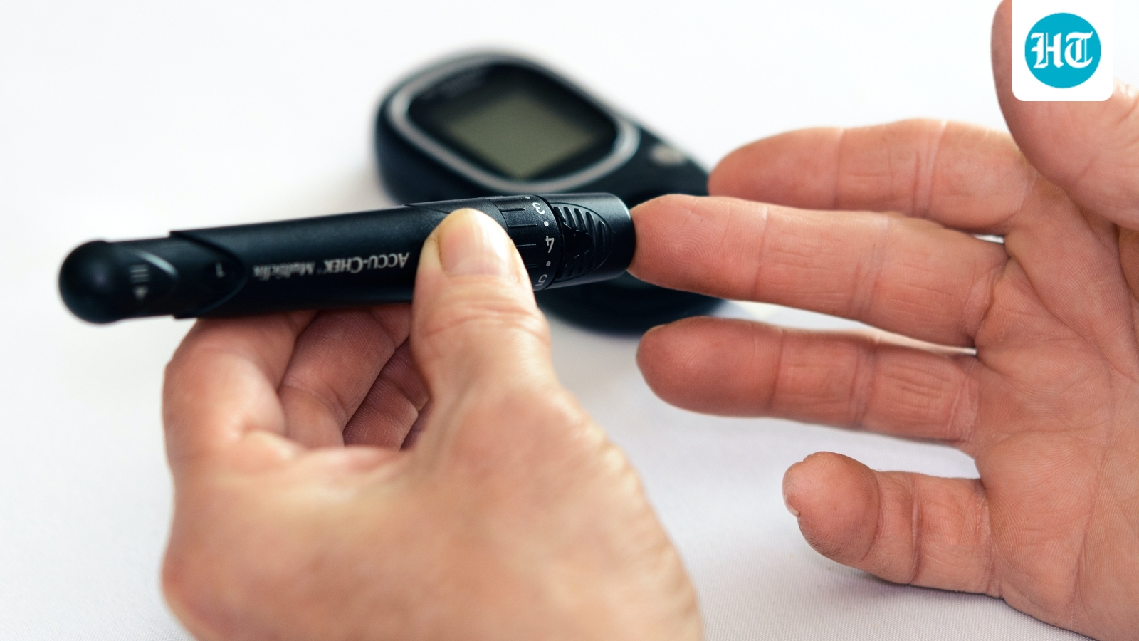
Category: 8. Health

  • South Africa’s HIV/Aids patients worry about treatment

    South Africa’s HIV/Aids patients worry about treatment

    Mayeni Jones

    BBC News, Johannesburg

    Reuters A glove-wearing nurse takes a blood sample from a child for an HIV test while the child's mother looks on at a clinic in Diepsloot, north of Johannesburg, South Africa, 12 March2025Reuters

    Gugu used to collect her anti-retroviral from a USAID-funded clinic in downtown Johannesburg.

    But when President Trump’s cuts to aid funding were announced earlier this year, her and thousands of other HIV-positive patients across South Africa suddenly faced an uncertain future.

    Gugu was lucky, the clinic where she got the medication that helps suppress her symptoms contacted her before it closed down.

    “I was one of the people who was able to get their medication in bulk. I usually collect a three-month prescription. But before my clinic closed, they gave me nine months’ worth of medication.”

    She will run out of ARVs in September, and then plans on going to her local public hospital for more.

    A former sex worker, the 54-year-old found out she was HIV-positive after she’d quit the industry.

    Ten years ago she got a chesty cough, and initially thought it was tuberculosis. She went to a doctor who told her she had a chest infection and treated her for it.

    But when the treatment failed, she went to a clinic to get an HIV test.

    “By then I already assumed that I was HIV-positive, and I told the nurse this.”

    She was right, and she has been on antiretrovirals (ARVs) ever since. We’re not using her real name at her request.

    She currently works as a project coordinator for an NGO.

    “We help pregnant sex workers get their ARVs, to ensure their children are born HIV-negative. We also do home visits to make sure that the mothers take their medication on time, and to look after their babies when they go for their monthly check-ups.”

    Many HIV-positive sex workers in South Africa relied on private clinics funded by the US government’s now-defunct aid agency, USAID, to get their prescriptions and treatments.

    But most of the facilities closed after US President Donald Trump cut most foreign aid earlier this year.

    In a report due to be released on Thursday, the UN body in charge of fighting HIV/Aids does not single out the US, but says that drastic cuts from a number of donors have sent shockwaves around the world, and the “phenomenal progress” in tackling the illness risks being reversed.

    “New HIV infections have been reduced by 40% since 2010, and 4.4 million children have been protected from acquiring HIV since 2000. More than 26 million lives have been saved,” UNAIDS says, warning that if the world does not act, there could be an extra six million new HIV infections and four million AIDS-related deaths by 2029.

    Gugu has so far been lucky. The clinic from where she got her antiretrovirals in Johannesburg contacted her before it closed.

    She will run out of antiretrovirals in September, and will then go to her local public hospital for more.

    She believes that many sex workers could be discouraged from doing so.

    “The problem with going to public hospitals is the time factor. In order to get serviced at these facilities, you have to arrive at 4 or 5am, and they may spend the whole day waiting for their medication. For sex workers, time is money,” Gugu says.

    She adds that she recently went to her local clinic with some friends to register her details and build a relationship with staff.

    “The nurse who attended to us was very rude. She told us there was nothing special about sex workers.”

    She thinks this could lead to many sex workers defaulting on their medication, “especially because their hospital files contain a lot of personal information, and the concern is that sometimes the nurses at these local clinics aren’t always the most sensitive in dealing with this kind of information.”

    According to the UN, the US cuts to HIV funding could reverse some of the gains made by what has been called one of the most successful public health interventions in history.

    Scientists in the UK-based Lancet medical journal last month estimated that USAID funding directly reduced Aids deaths by 65%, or 25.5 million, over the past two decades.

    Getty Images Standing at a podium, George W. Bush, in a suit, turns his head as South Africa's Thandazile Darby and Dr Helga Holst, both seated with children, applaud on 1 December 2005 as World Aids Day is commemorated in the Eisenhower Executive Office Building in Washington, DC.Getty Images

    Former US President George W Bush is widely acknowledged for his commitment to tackling HIV/Aids

    Then-US President George W Bush launched an ambitious programme to combat HIV/Aids in 2003, saying it would serve the “strategic and moral interests” of the US.

    Known as the President’s Emergency Plan for Aids Relief (Pepfar), it led to the investment of more than $100bn (£74bn) in the global HIV/Aids response – the largest commitment by any nation to address a single disease in the world.

    South Africa has about 7.7 million people living with HIV, the highest number in the world, according to UNAIDS.

    About 5.9 million of them receive antiretroviral treatment, resulting in a 66% decrease in Aids-related deaths since 2010, the UN agency adds.

    South Africa’s government says Pepfar funding contributed about 17% to its HIV/Aids programme. The money was used for various projects, including running mobile clinics to make it easier for patients to get treatment.

    The Trump administration’s cuts have raised concern that infection rates could spike again.

    “I think we’re going to start seeing an increase in the number of HIV infections, the number of TB cases, the number of other infectious diseases,” Prof Lynn Morris, Deputy Vice-Chancellor of Johannesburg’s Wits University, tells the BBC.

    “And we’re going to start seeing a reversal of what was essentially a real success story. We were getting on top of some of these things.”

    Gugu points out that treatment is a matter of life and death, especially for vulnerable populations like sex workers.

    “People don’t want to default on their ARVs. They’re scared that they’re going to die if they don’t get access to them.

    The cuts have also affected research aimed at finding an HIV vaccine and a cure for Aids.

    “There’s the long-term impact, which is that we’re not going to be getting new vaccines for HIV,” Prof Morris adds.

    “We’re not going to be keeping on top of viruses that are circulating. Even with new viruses that might appear, we’re not going to have the surveillance infrastructure that we once had.”

    South Africa has been one of the global leaders in HIV research. Many of the medications that help prevent the virus, and which have benefitted people around the world, were trialled in South Africa.

    This includes Prep (pre-exposure prophylaxis), a medication which stops HIV-negative people from catching the virus.

    Another breakthrough preventive drug released this year, Lenacapavir, an injection taken twice a year and that offers total protection from HIV, was also tried in South Africa.

    Prof Abdullah Ely is in his lab, in a white coat and blue gloves

    South African academic Prof Abdullah Ely is concerned that research will be affected by the US funding cut

    In a lab at Wits University’s Health Sciences campus, a small group of scientists are still working on a vaccine for HIV.

    They are part of the Brilliant Consortium, a group of labs working across eight African countries to develop a vaccine for the virus.

    “We were developing a vaccine test to see how well that works, and then we would trial it on humans,” Abdullah Ely, an Associate Professor at Wits University, tells the BBC in his lab.

    “The plan was to run the trials in Africa based on research carried out by Africans because we want that research to actually benefit our community as well as all mankind.”

    But the US funding cuts threw their work into doubt.

    “When the stop order came, it meant we had to stop everything. Only some of us have been able to get additional funding so we could continue our work. It’s set us back months, probably could even be a year,” Prof Ely says.

    The lab lacks funding to carry out clinical trials scheduled for later this year.

    “That is a very big loss to South Africa and the continent. It means that any potential research that comes out of Africa will have to be tested in Europe, or the US,” Prof Ely says.

    In June, universities asked the government for a bailout of 4.6bn South African rand ($260m; £190m) over the next three years to cover some of the funding lost from the US.

    “We are pleading for support because South Africa is leading in HIV research, but it’s not leading for itself. This has ramifications on the practice and policies of the entire globe,” says Dr Phethiwe Matutu, head of Universities South Africa.

    South Africa’s Health Minister Aaron Motsoaledi announced on Wednesday that some alternative funding for research had been secured.

    The Bill and Melinda Gates Foundation and the Wellcome Trust have agreed to donate 1m rand each with immediate effect, while the government would make available 400m rand over the next three years, he said.

    This would bring the total to 600m rand, way below the 4.6bn rand requested by researchers.

    As for Gugu, she had hoped that by the time she was elderly, a cure for HIV/Aids would have been found, but she is less optimistic now.

    “I look after a nine-year-old. I want to live as long as I can to keep taking care of him,” she tells the BBC.

    “This isn’t just a problem for right now, we have to think about how it’s going to affect the next generation of women and young people.”

    You may also be interested in:

    Getty Images/BBC A woman looking at her mobile phone and the graphic BBC News AfricaGetty Images/BBC

    Continue Reading

  • New analysis shows stopping antidepressants rarely leads to severe symptoms

    New analysis shows stopping antidepressants rarely leads to severe symptoms

    The largest review of ‘gold standard’ antidepressant withdrawal studies to date has identified the type and incidence of symptoms experienced by people discontinuing antidepressants, finding most people do not experience severe withdrawal.

    In a systematic review and meta-analysis of previous randomised controlled trials relating to antidepressant withdrawal, a team of researchers led by Imperial College London and King’s College London concluded that, while participants who stopped antidepressants did experience an average of one more symptom than those who continued or were taking placebos, this was not enough to be judged as significant.

    The most common symptoms were dizziness, nausea, vertigo and nervousness. Importantly, depression was not a symptom of withdrawal from antidepressants, and was more likely to reflect illness recurrence.

    Researchers at Imperial College London, King’s College London, UCL and UK collaborators say their study provides much needed, clearer guidance for clinicians, patients and policymakers.

    Dr. Sameer Jauhar, lead author, at Imperial College London, said: “Our work should reassure the public because we replicated other findings, from high-quality studies, and have highlighted the clinical symptoms to look out for. Despite previous concern about stopping antidepressants, our work finds that most people do not experience severe withdrawal, in terms of additional symptoms. Importantly, depression relapse was not linked to antidepressant withdrawal in these studies, suggesting that if this does occur, people will need to see their health professional to rule out a recurrence of their depressive illness.”

    Clinical academics from around the UK worked collaboratively to conduct the largest and most rigorous analysis of randomised controlled trials in antidepressant withdrawal, examining data from 50 trials across multiple conditions. The data involved a total of 17,828 participants, with an average age of 44 years, of whom 70% were female. Two meta-analyses were conducted, one of the trials that used a standardised measure known as the Discontinuation Emergent Signs and Symptoms scale (DESS), and the other of the trials that used various other scales.

    Across antidepressants, irrespective of type taken, the number of extra symptoms generally equated to one more symptom on the 43-symptom item scale. In placebo-controlled randomised controlled trials, the most common symptoms across antidepressants were dizziness (7.5% vs 1.8%), nausea (4.1% vs 1.5%), vertigo (2.7% vs 0.4%) and nervousness (3% vs 0.8%).

    Experiencing just one symptom is below the 4 or more cutoff for clinically important discontinuation syndrome. 

    The nature, and rates, of different symptoms varied between antidepressants, and some symptoms were also seen with placebo. This helped to clarify which symptoms were likely to be illness recurring, such as the participant relapsing into depression.

    The data involved different types of antidepressants, including the serotonin-norepinephrine reuptake inhibitors (SNRIs) venlafaxine and duloxetine; the selective serotonin reuptake inhibitors escitalopram, sertraline and paroxetine; agomelatine, which is a melatonin receptor agonist and selective serotonin receptor antagonist; and vortioxetine, which inhibits the reuptake of serotonin as well as partial agonist and antagonist effects on various serotonin receptors.

    The most symptoms were seen with discontinuance of venlafaxine, where approximately 20% of people suffered from dizziness, compared to 1.8% taking placebo. With vortioxetine, fewer than one extra symptom was seen on the standardised discontinuation scale. No extra symptoms were seen with agomelatine.

    Adding non-placebo controlled studies increased these rates slightly; dizziness (11.8%, nightmares 8.1%, nervousness 7.6%, nausea 5.8%).

    Relapse of depression was not seen in those withdrawing from antidepressants, even in people with existing depression.

    The review included studies with different discontinuation regimes, but in the majority of studies (44), people either discontinued abruptly or tapered over 1 week.

    While uncommon, our study highlights that there could be a sub-group of people who develop more severe withdrawal symptoms than the wider population of antidepressant users. Our focus must now turn to look at the pharmacological basis for this reaction, and ask whether it relates to the way they metabolise these drugs.”


    Michail Kalfas, of the Institute of Psychiatry, Psychology & Neuroscience at King’s College London

    In terms of study limitations, 38 of the trials followed people up for up to two weeks post-discontinuation (the time period one would expect most discontinuation symptoms to occur), so researchers say this limits long-term conclusions. However, they note that findings from the 2021 UCL-led ANTLER trial involving long-term antidepressant users – which was included in this review – suggested severe withdrawal is infrequent, even after prolonged use.

    The study follows recent concerns about the effects of stopping antidepressants, as well as various guidance changes on their prescribing. This current meta-analysis helps resolve the debate by showing that withdrawal is a real and drug-specific phenomenon, though not an inevitable outcome.

    Professor Allan Young, Head of Psychiatry at the Department of Brain Sciences at Imperial College London, said: “Depression and anxiety are common conditions and antidepressant treatments are effective and generally quite well tolerated. However, concerns have been raised about the after-effects of stopping these treatments and this is something that has affected patients and clinicians. Changes of guidance may also have impacted the use of these treatments. Now, this cutting-edge review clarifies the scientific evidence and should reassure all parties about the use, and discontinuation, of these treatments. Official guidance should now be changed to reflect the evidence.”

    Incidence and Nature of Antidepressant Discontinuation Symptoms, A Systematic Review and Meta-analysis by Michail Kalfas, Sameer Jauhar et al is published in JAMA Psychiatry on 9th July 2025. DOI: 10.1001/jamapsychiatry.2025.1362.

    The authors will present their findings and discuss the clinical implications in a briefing hosted by the Science Media Centre online on Tuesday 8th July at 10.30am UK time. For an invitation, please contact Freya Robb, Science Media Centre: [email protected]

    Source:

    Journal reference:

    Kalfas, M., et al. (2025). Incidence and Nature of Antidepressant Discontinuation Symptoms. JAMA Psychiatry. doi.org/10.1001/jamapsychiatry.2025.1362.

    Continue Reading

  • Interaction between insulin resistance and systemic inflammation on ca

    Interaction between insulin resistance and systemic inflammation on ca

    Introduction

    Coronary artery disease (CAD) remained the primary cause of mortality and posed a growing public health challenge globally.1,2 Coronary artery bypass grafting (CABG) served as the cornerstone of CAD management and was preferred for patients with complex CAD, left main disease, or multi-vessel disease.3,4 However, the long-term prognosis post-CABG remained suboptimal. Recent studies demonstrated that bypass grafts had high failure rates, with 3.6% occluding at 5 years and approximately 11% failing at 8 years after CABG.5,6 Consequently, the early identification of high-risk patients and management of pertinent risk factors were critical for improving postoperative outcomes.

    Insulin resistance (IR), characterized by diminished responsiveness of target tissues to insulin stimulation,7 was not only recognized as an independent risk factor for atherosclerotic cardiovascular disease8,9 but also associated with elevated susceptibility to adverse cardiovascular events.10 The estimated glucose disposal rate (eGDR), a novel non-insulin-based surrogate marker for IR, integrated blood pressure and waist circumference rather than relying solely on fasting glucose and insulin levels.11,12 This approach provided a more comprehensive assessment of metabolic health compared to traditional measures like glycated hemoglobin (HbA1c)12 or homeostasis model assessment of insulin resistance (HOMA-IR).13 Prior studies indicated that eGDR exhibited stronger predictive power for cardiovascular risk than HbA1c14 or HOMA-IR,13 particularly in non-diabetic populations.15–17 Nevertheless, the prognostic value of eGDR in CAD patients undergoing CABG remained unclear.

    Inflammatory response played a pivotal role in CAD pathogenesis.18 C-reactive protein (CRP), a classic inflammatory biomarker, was confirmed as an independent CAD risk factor, with predictive utility comparable to lipid profiles or blood pressure.18,19 Both the Centers for Disease Control and Prevention and the American Heart Association (AHA) recommended CRP > 3.0 mg/L as a high-risk threshold for guiding cardiovascular risk stratification.20,21 Importantly, IR was frequently associated with elevated CRP levels in prior studies,22,23 suggesting potential mechanistic interplay. However, the combined impact of IR and inflammation on cardiovascular outcomes after CABG had not been systematically investigated.

    Therefore, in the present study, we sought to investigate the relationship between IR assessed by the eGDR and the inflammation evaluated by CRP levels with long-term cardiovascular outcomes in non-diabetic patients undergoing CABG. Specifically, 1) to evaluate the relationship between eGDR and CRP in non-diabetic individuals undergoing CABG. 2) to examine whether eGDR and CRP have a synergistic effect in predicting the prognosis of CABG patients; 3) to assess whether inflammation mediates the relationship between IR and cardiovascular outcomes.

    Methods

    Study Population

    A total of 1658 patients diagnosed with CAD and undergoing CABG surgery were consecutively enrolled at the Second Xiangya Hospital of Central South University (Changsha, China) from April 2011 to December 2020. The diagnosis of CAD was based on typical angina pectoris, and severe stenosis of ≥1 of the coronary arteries, as indicated by coronary angiography. The exclusion criteria were as follows: 1) a history of diabetes; 2) those aged ≥80 years; 3) patients who underwent aortic valve replacement, mitral valve replacement, aortic root replacement surgery for aortic root aneurysm and aortic dissection, or complex congenital heart disease surgery at the same time; 4) those who died during index hospitalization or within 1 year after discharge; 5) and those lost to follow-up or with missing baseline or follow-up data. This study was approved by the ethics committee of the Second Xiangya Hospital of Central South University and strictly complied with the Declaration of Helsinki, and informed consent was waived due to the retrospective nature of the study and the anonymized processing of patient data.

    Data Collection and Definitions

    All anthropometric parameters, clinical history, and laboratory test results were acquired from electronic medical records. The anthropometric characteristics included age, sex, body mass index (BMI), waist circumference (WC) and smoking status. The clinical history included hypertension, chronic kidney disease (CKD), diabetes, prior percutaneous coronary intervention (PCI), myocardial infarction (MI) ACS/CCS prevalence and number and types of grafts. Antiplatelet, lipid-lowering, antihypertensive, and antidiabetic medications have also been recorded.

    Fasting venous blood samples were collected to measure the plasma levels of hemoglobin, glycosylated hemoglobin A1c (HbA1c), serum creatinine (Scr), total cholesterol (TC), triglycerides (TG), low-density lipoprotein cholesterol (LDL-C), high-density lipoprotein cholesterol (HDL-C), and serum creatinine employing standard laboratory methods. The left ventricular ejection fraction (LVEF) was measured using the two-dimensional modified Simpson’s method.

    The diagnosis of CAD was based on typical angina pectoris, and severe stenosis of ≥ 1 of the coronary arteries, as indicated by coronary angiography.24 The identification of diabetes was based on either the self-reported use of antidiabetic medications or elevated blood glucose readings, characterized by casual blood glucose levels of 11.1 mmol/L or higher, fasting blood glucose levels of 7.0 mmol/L or higher, or 2-hour postprandial levels exceeding 11.1 mmol/L following a 75 g oral glucose tolerance test.25 Hypertension was identified through a consistent record of blood pressure readings of 140/90 mmHg or above, or the ongoing use of antihypertensive medication.26

    eGDR was determined using the formula: 21.158 – (0.09 × waist circumference [cm]) − (3.407 × hypertension [yes 1 or no 0]) − (0.551 × glycated hemoglobin A1c [HbA1c] [%]).12

    Follow-up and Endpoints

    The clinical follow‐up data after CABG were collected by reviewing serial records of patients who visited outpatient clinics and telephone interviews with all other patients. The primary end point was major adverse cardiovascular and cerebrovascular events (MACCEs), which were defined as a composite of cardiac death, nonfatal MI, any revascularization, including native coronary arteries and bypass grafts (internal mammary artery, radial artery, and saphenous vein grafts), cardiac rehospitalization (admission because of angina or heart failure), and nonfatal stroke.

    Statistical Analysis

    Continuous variables were characterized by the mean ± SD or the median with interquartile range (IQR), based on the data’s distribution normality. Group differences were analyzed using t-tests or Mann–Whitney U-tests accordingly. Categorical variables were summarized as frequencies and percentages, and comparisons between groups were performed using the chi-square (χ2) test or Fisher’s exact test. Spearman coefficients and linear regression models were performed to assess the association between eGDR and CRP. The incidence of MACCEs in different groups was assessed by the Kaplan–Meier method based on the eGDR or CRP groups, respectively.

    The dose-response association between the eGDR, CRP, and MACCEs in patients after CABG was illustrated through restricted cubic splines (RCS) curve. Cox regression models were employed to assess the relationship between the eGDR, CRP, and the incident MACCEs. Receiver operating characteristic (ROC) curves and the area under the curves (AUC) were constructed to compare the predictive value of eGDR and CRP for MACCEs. Additionally, C-statistics, a net reclassification index (NRI), and an integrated discrimination improvement (IDI) to evaluate the incremental predictive value of the individual and combined eGDR and CRP. Subgroup analyses were conducted to explore whether the predictive utility of the eGDR and CRP remained consistent across patients with diverse demographic characteristics or comorbidities. In mediation analysis, we employed VanderWeele’s two-stage regression method to obtain survival data. Specifically, we utilized Cox proportional hazards regression to analyze the outcome (MACCEs) and linear regression for the mediator (CRP), evaluating the significance of the mediating effect through examination of 1000 bootstrap samples.

    In liner and COX regression analysis and mediation analysis, we employed multiple adjusted models, adjusting various covariates independently, to thoroughly evaluate the robustness and reliability of the findings. Model I was adjusted for age and sex. Model II was adjusted for age, sex, CKD, LVEF, smoker, previous MI, previous PCI and SYNTAX scores. Model III was adjusted for age, sex, CKD, LVEF, smoker, previous MI, previous PCI, SYNTAX scores, statins, Aspirin, P2Y12 inhibitors, ACEI/ARB, β-blockers, TC, LDL-C, HDL-C, TG and Scr. The associations were further assessed with the inverse probability of censoring weighted (IPCW) method as a sensitivity analysis.27 In the IPCW model, the probability for predicting complete data was generated based on all non-diabetic patients undergoing CABG regardless of missing data. The results were considered statistically significant when the 2-sided P value was <0.05. R version 4.0.5 (R Foundation for Statistical Computing) in RStudio version 1.1.463 (RStudio, Inc) and Prism version 8.0.2 were used to perform all statistical analyses.

    Results

    Baseline Characteristics

    The final cohort consisted of 1658 participants who were eligible for the final analysis (aged 60.8 ± 8.3 years; 76.7% men). Over a median follow-up period of 60.9 months, 414 MACCEs cases were observed. Baseline characteristics of the study population are presented in Table 1. No significant differences were observed in sex distribution, ACS/CCS prevalence, diabetes markers HbA1c, smoking status, renal function parameters, or perioperative medication use (all P > 0.05). Surgical characteristics including graft numbers and conduit types showed comparable distributions between groups.

    Table 1 Baseline Characteristics of Participants Stratified by the Occurrence of MACCEs

    Patients who experienced MACCEs were generally older and exhibited higher levels of BMI, WC, TC, LDL-C, TG, CRP and SYNTAX score and incidence of hypertension (all P < 0.05). They also had significantly lower LVEF, eGDR index and HDL-C level (all P < 0.05).

    Association Between eGDR and CRP

    Patients were divided into Tertile 1 group (eGDR ≤ 7.36), Tertile 2 group (7.36 < eGDR ≤ 8.78) and Tertile 3 group (eGDR > 8.78) according to eGDR tertiles. The CRP levels decreased with increasing tertiles of eGDR index (Figure 1A). CRP levels were negatively associated with eGDR index (R = -0.45, P < 2.2e−16; Figure 1B). Additionally, regarding linear regression models measuring eGDR as a continuous variable, each SD increment in eGDR was associated with a 0.51 mg/L decrease in CRP levels (95% CI: −0.61– −0.40; P < 0.001) after adjusting for all covariates (Table S1). Likewise, the categorical analysis revealed that, compared with the Tertile1 group, the Tertile3 group was significantly associated with a 1.72 mg/L (95% CI: −2.15 – −1.29; P < 0.001) decrease (Table S1).

    Figure 1 Association between eGDR and CRP. (A) Violin plot showing the distribution of CRP among groups categorized by eGDR tertiles; (B) scatter plot.

    Abbreviations: eGDR, estimated glucose disposal rate; CRP, C-reactive protein.

    Association Between the eGDR Index, CRP Levels, and the Incident MACCEs

    Patients were further divided into Tertile1 group (CRP ≤ 2.28 mg/L), Tertile 2 group (2.28 < CRP ≤ 3.87 mg/L) and Tertile 3 group (CRP > 3.87 mg/L) according to CRP tertiles. Kaplan–Meier survival curves of eGDR and CRP for long-term MACCEs are plotted in Figure 2. The MACCEs incidence increased with increasing tertile of the CRP levels and decreasing tertile of eGDR (all log-rank P < 0.001). When analyzed as continuous variables, an decreased eGDR (HR: 0.792, 95% CI: 0.749–0.875, P < 0.001; Table 2) and increased CRP levels (HR: 1.042, 95% CI: 1.029–1.054, P < 0.001; Table S2) were independently associated with MACCEs in the fully adjusted model. Similarly, when analyzed as categorical variables, the Tertile 3 group of eGDR exhibited a lower incidence (HR: 0.523, 95% CI: 0.409–0.668, P < 0.001; Table 2), whereas the Tertile 3 group of CRP showed a higher incidence of MACCEs (HR: 1.747, 95% CI: 1.364–2.238, P < 0.001; Table S2). In the sensitivity analysis considering the potential bias due to missing data of the eGDR or CRP with the IPCW method, the association between the eGDR or CRP and MACCEs remained unchanged (IPCW model in Table 2 and Table S2).

    Table 2 Association of the eGDR with the Risk of MACCEs in Non-Diabetic Patients Undergoing CABG

    Figure 2 Cumulative incidence of MACCEs during follow-up stratified by the eGDR (A) and CRP levels (B).

    Abbreviations: eGDR, estimated glucose disposal rate; CRP, C-reactive protein; MACCEs, major adverse cardiovascular and cerebrovascular events.

    Subgroup analyses were conducted to assess whether the predictive value of the eGDR and CRP remained consistent across diverse demographic characteristics or comorbidities. After stratifying by sex, age, BMI, hypertension, smoking status and ACS/CCS distribution, both decreased eGDR (Figure S1) and elevated CRP (Figure S2) emerged as significant predictors of MACCEs across various subgroups.

    Synergistic Effect of eGDR and CRP on Prediction of Incident MACCEs

    According to the RCS analyses, the association between eGDR and MACCEs followed a L-shape and the risk of MACCEs significantly increased when eGDR was lower than 8 mg/kg/min (Figure 3A). While a positive dose–response relationship between the CRP and MACCEs was observed (Figure 3B). To evaluate the addictive effect of eGDR and CRP in predicting MACCEs, the patients were re-categorized by a combination of eGDR (ie 8 mg/kg/min) based on the values obtained by RCS and CRP according to AHA (ie 3 mg/L).21 In comparison with the group with eGDR > 8 and CRP < 3, the group with eGDR ≤ 8 and CRP ≥ 3 had approximately 2.28 times the risk of incident MACCEs (HR: 2.282, 95% CI: 1.749–2.978, P < 0.001; Table 3) after adjusting for all covariates. Sensitivity analysis suggested that the synergistic effect remained unchanged (IPCW model in Table 3). ROC curves of eGDR, CRP, and their combination are plotted in Figure 4. The combination of eGDR and CRP performed better than eGDR (0.645 vs 0.624, P = 0.0382) and CRP (0.645 vs 0.612, P = 0.0251) alone in predicting incident MACCEs. Furthermore, C-statistics, NRI, and IDI were analyzed. Adding eGDR and CRP to the basic model simultaneously further improved C-statistics (0.626, 95% CI: 0.604-0.645 vs 0.556, 95% CI:0.539–0.573, P < 0.001). Additionally, the risk reclassification and discriminatory power also appeared to be substantially better, with an IDI of 0.023 (95% CI: 0.011–0.037; P < 0.001), and a NRI of 0.036 (95% CI: 0.021–0.071; P < 0.001) (Table S3). These findings indicated that combining eGDR and CRP improved the prediction efficiency for MACCEs.

    Table 3 Association Between the Combination of the eGDR and CRP and MACCEs in Non-Diabetic Patients Undergoing CABG

    Figure 3 Dose-responsive relationship of eGDR (A) and CRP (B) with the risk of with MACCEs in non-diabetic individuals undergoing CABG.

    Abbreviations: eGDR, estimated glucose disposal rate; CRP, C-reactive protein; MACCEs, major adverse cardiovascular and cerebrovascular events; CABG, coronary artery bypass grafting; HR, hazard ratio; CI, confidence interval.

    Figure 4 Receiver operating characteristic curves of eGDR, CRP, and their combination in predicting incident MACCEs.

    Abbreviations: eGDR, estimated glucose disposal rate; CRP, C-reactive protein; MACCEs, major adverse cardiovascular and cerebrovascular events; CABG, coronary artery bypass grafting; AUC, area under curve.

    Mediating Effects of CRP on the Association Between eGDR and Incident MACCEs

    As demonstrated in Tables 4 and 5, the mediation analysis revealed that the CRP levels, exerted a significant partial mediating effect on the relationship between IR, as reflected by the eGDR, and the incidence of MACCEs across multiple adjusted models. Specifically, the mediation proportions of an elevated CRP levels were 11.8% (CI: 5.6–17.9%, P < 0.001), 12.8% (CI: 3.0–22.6%, P = 0.011), 12.5% (CI: 2.6–22.4%, P = 0.017) and 12.3% (CI: 3.8–20.8%, P = 0.004) in the crude, adjusted Model I, adjusted Model II, and adjusted Model III, respectively.

    Table 4 Decomposition of the Total Association of the eGDR and the Risk of MACCEs in Nondiabetic Patients Undergoing CABG Into Direct and Indirect Associations Mediated by Baseline CRP

    Table 5 Decomposition of the Total Association of the eGDR and the Risk of MACCEs in Non-Diabetic Patients Undergoing CABG Into Direct and Indirect Associations Mediated by Baseline CRP

    Discussion

    The principal findings of the present investigation were as follows: 1) eGDR was negatively correlated with plasma CRP levels. 2) A significant association was found between decreased eGDR, elevated CRP levels and a higher incidence of MACCEs post CABG, which remained in different models, sensitivity and subgroup analyses. 3) There was a potential synergistic effect of eGDR and CRP on MACCEs. The combination of eGDR ≤ 8 and CRP ≥ 3 can effectively identify individuals at the highest risk of MACCEs undergoing CABG. 4) Increased CRP levels partly mediated the connection between eGDR and MACCEs, in non-diabetic patient following CABG.

    IR is a metabolic disorder significantly related to the occurrence and development of atherosclerotic cardiovascular disease. The current gold standard for analyzing IR is the hyperinsulinemic-euglycemic clamp,28 but it is not suitable for clinical practice and large cohort studies due to its invasiveness and cost. Previous studies have defined insulin resistance using the HOMA-IR index, which is calculated based on fasting glucose and fasting insulin.29 However, routine measurement of fasting insulin levels is not common in standard clinical management of CABG, especially in non-diabetic patients undergoing CABG. eGDR, based on patient’s body size, HbA1c, and presence of hypertension, all of which are included in routine assessments of CAD patients upon hospital admission, is more suitable for secondary prevention in patients following CABG. Sun et al discovered a correlation between eGDR and arterial stiffness and found that it could predict long-term all-cause mortality.30 A large-scale population study emphasized that eGDR can enhance the diagnostic accuracy of ischemic heart disease in the general population.31 In a retrospective study involving non-diabetic patients with non-ST-segment elevation acute coronary syndrome, the findings indicated that low eGDR were a significant risk factor for adverse cardiovascular events.17 Consistent with existing research, this present study illustrated an independent association between a lower eGDR and MACCEs in different models and subgroup analyses in non-diabetic patients after CABG. There was a L-shaped relationship between eGDR and mortality, with cutoff values > 8, which is similar to the cut-values recommended by previous studies for eGDR (<4, 4–6, 6–8, and ≥ 8mg/kg/min).12,32

    Atherosclerotic cardiovascular disease has been characterized as a chronic inflammatory condition, highlighting the significant role of inflammation in its pathogenesis and progression. CRP is a systemic inflammatory marker, which activates multiple processes of atherosclerosis, including but not limited to monocyte cytokine expression, adhesion molecule expression, and platelet aggregation.33 Prior studies have demonstrated that the serum CRP levels are higher in patients with acute myocardial infarction patients compared to those with stable angina patient, and CAD patients with higher concentrations of CRP have poorer cardiovascular prognosis.34,35 In line with these findings, an significantly positive association between CRP levels and MACCEs in different models and subgroup analyses was observed. While CRP served as a pragmatic marker of systemic inflammation in this cohort, emerging biomarkers like growth differentiation factor-15 (GDF-15)36 and lipoprotein-associated phospholipase A2 (Lp-PLA2),37 which directly reflect plaque vulnerability and vascular stress-merit investigation in future studies to unravel tissue-specific inflammatory mechanisms underlying IR-driven cardiovascular risk.

    IR plays a crucial role as a potential mechanism for increasing CVD risk by activating inflammation-related genes and lead to chronic inflammation, thereby impairing vascular health and promoting CVD.38 A cross-sectional study showed that serum high sensitive CRP was positively correlated with HbA1c and HOMA-IR in patients with subclinical atherosclerosis.39 Similarly, we found a significantly negative correlation between eGDR and CRP in non-diabetic individuals following CABG.

    Additionally, in our study, we observed the synergistic effects of eGDR and CRP on long-term MACCEs. Combining them may help further risk stratification for non-diabetic individuals undergoing CABG. The ROC curve demonstrated that the predicting ability for MACCEs using a combination of the eGDR and CRP is greater than that of either index alone. This phenomenon arises from the eGDR index primarily evaluating insulin resistance levels, while CRP reflects factors such as inflammatory burden, thereby resulting in a complementary effect. Consistent with our findings, Li et al reported that IR and systemic inflammation synergistically increase the risk of cardiovascular events in patients with chronic coronary syndrome.40

    More importantly, this study clarified the mediating role of CRP levels in linking the eGDR to MACCEs, consolidating previous findings into a comprehensive pathway to guide clinical decision-making. To our knowledge, this study is the first evidence on the causal pathways of insulin resistance, inflammatory markers, and cardiovascular adverse outcomes in non-diabetic patients undergoing CABG. Despite the precise mechanism of mediation interaction remaining unclear, some previous studies provided valuable mechanistic insights. Prior studies suggested that systemic inflammation partially mediates the association between IR and clinical outcomes.40,41 These observational findings provide epidemiological evidence supporting the biologically plausible notion that inflammation could serve as a mediator in the association between IR and adverse outcomes.42 Mechanistically, IR can activate the NOD-like receptor protein 3 inflammasome, which is a key component in the pathogenesis of atherosclerosis, and CRP is a downstream marker.43 Therefore, IR, represented by decreased eGDR, might exacerbate atherosclerosis by up-regulating the NLRP3 signaling pathway, represented by CRP. Additional research is warranted to investigate the potential mechanisms that underlie the causal relationship between eGDR and cardiovascular events. Clinical trials have demonstrated the potential of anti-inflammatory therapy in improving cardiovascular outcomes in secondary prevention of CAD.44,45 However, incorporating anti-inflammatory therapy into CAD management still poses challenges. One obstacle is the need for more precise risk stratification to enhancing cost-effectiveness. This study proposes that utilizing eGDR and CRP in combination can aid in identifying patients at significantly elevated cardiovascular risk. Furthermore, the relationship between low eGDR and adverse cardiovascular outcomes may be mediated by CRP. In addition to directly reducing systemic inflammation, anti-inflammatory treatment targeting these individuals may offer additional benefits, including reducing the synergistic and mediating effects of inflammation on the adverse outcomes of IR.

    Limitation

    Although offering valuable insights, this study also presents limitations that require thoughtful consideration. First, exclusion criteria for the study included patients without data for the eGDR and CRP, potentially introducing selection bias. Nevertheless, sensitivity analyses utilizing IPCW methods to address missing data yielded results consistent with the primary findings. Second, the limitation of a single-center observational design hinders our ability to establish causal relationships between the eGDR, CRP levels, and MACCEs post-CABG. Third, while many important confounding factors were considered in the multivariable analysis, the impact of unmeasured or unknown confounders on outcomes cannot be entirely ruled out. Forth, the eGDR and CRP data were mostly obtained upon patient admission, thus we were unable to ascertain the impact of dynamic changes in eGDR and CRP on prognosis. Finally, although our study reveals an inverse association between eGDR and CRP levels, future human studies using direct IR measurements (eg, hyperinsulinemic-euglycemic clamps) and other inflammatory marker assessments (eg, GDF-15, Lp-PLA2, interleukin-6) are needed to determine whether IR drives inflammation.

    Data Sharing Statement

    The dataset analyzed during the current study is available from the corresponding author on reasonable request.

    Ethics Approval and Consent to Participate

    The study was approved by the ethics committee of Second Xiangya Hospital of Central South University, with a waiver of informed consent.

    Acknowledgments

    Yingying Xie and Hao Chen are co-first authors for this study. The authors would like to acknowledge the patient participants and their relatives, the clinical and research teams, and the nursing teams at all hospitals for their contribution to the study without financial compensation.

    Funding

    This work was supported by National High Level Hospital Clinical Research Funding (2024-NHLHCRF-YS-01). National Natural Science Foundation of China (No. 82270352). National High Level Hospital Clinical Research Funding (2024-NHLHCRF-JBGS-WZ-06). National Key Clinical Specialty Construction Project (No. 2020-QTL-009). National High Level Hospital Clinical Research Funding (2023-NHLHCRF-YXHZ-ZRMS-09).

    Disclosure

    The authors report no conflicts of interest in this work.

    References

    1. Virani SS, Alonso A, Benjamin EJ, et al. Heart disease and stroke statistics-2020 update: a report from the American Heart Association. Circulation. 2020;141(9):e139–e596. doi:10.1161/CIR.0000000000000757

    2. Vos T, Lim SS, Abbafati C, et.al. Global burden of 369 diseases and injuries in 204 countries and territories, 1990-2019: a systematic analysis for the global burden of disease study 2019. Lancet. 2020;396(10258):1204–1222. doi:10.1016/S0140-6736(20)30925-9

    3. Neumann FJ, Sousa-Uva M, Ahlsson A, et al. 2018 ESC/EACTS Guidelines on myocardial revascularization. Eur Heart J. 2019;40(2):87–165. doi:10.1093/eurheartj/ehy394

    4. Mohr FW, Morice M-C, Kappetein AP, et al. Coronary artery bypass graft surgery versus percutaneous coronary intervention in patients with three-vessel disease and left main coronary disease: 5-year follow-up of the randomised, clinical SYNTAX trial. Lancet. 2013;381(9867):629–638. doi:10.1016/S0140-6736(13)60141-5

    5. Kim MS, Hwang HY, Kim JS, et al. Saphenous vein versus right internal thoracic artery as a Y-composite graft: five-year angiographic and clinical results of a randomized trial. J Thorac Cardiovasc Surg. 2018;156(4):1424–1433.e1. doi:10.1016/j.jtcvs.2018.04.123

    6. Xenogiannis I, Zenati M, Bhatt DL, et al. Saphenous vein graft failure: from pathophysiology to prevention and treatment strategies. Circulation. 2021;144(9):728–745. doi:10.1161/CIRCULATIONAHA.120.052163

    7. Defronzo RA, Lecture B. From the triumvirate to the ominous octet: a new paradigm for the treatment of type 2 diabetes mellitus. Diabetes. 2009;58(4):773–795. doi:10.2337/db09-9028

    8. Devesa A, Fuster V, Vazirani R, et al. Cardiac insulin resistance in subjects with metabolic syndrome traits and early subclinical atherosclerosis. Diabetes Care. 2023;46(11):2050–2057. doi:10.2337/dc23-0871

    9. Iglesies-Grau J, Garcia-Alvarez A, Oliva B, et al. Early insulin resistance in normoglycemic low-risk individuals is associated with subclinical atherosclerosis. Cardiovasc Diabetol. 2023;22(1):350. doi:10.1186/s12933-023-02090-1

    10. Laakso M, Kuusisto J. Insulin resistance and hyperglycaemia in cardiovascular disease development. Nat Rev Endocrinol. 2014;10(5):293–302. doi:10.1038/nrendo.2014.29

    11. Williams KV, Erbey JR, Becker D, et al. Can clinical factors estimate insulin resistance in type 1 diabetes? Diabetes. 2000;49(4):626–632. doi:10.2337/diabetes.49.4.626

    12. Zabala A, Darsalia V, Lind M, et al. Estimated glucose disposal rate and risk of stroke and mortality in type 2 diabetes: a nationwide cohort study. Cardiovasc Diabetol. 2021;20(1):202. doi:10.1186/s12933-021-01394-4

    13. Epstein EJ, Osman JL, Cohen HW, et al. Use of the estimated glucose disposal rate as a measure of insulin resistance in an urban multiethnic population with type 1 diabetes. Diabetes Care. 2013;36(8):2280–2285. doi:10.2337/dc12-1693

    14. Helliwell R, Warnes H, Kietsiriroje N, et al. Body mass index, estimated glucose disposal rate and vascular complications in type 1 diabetes: beyond glycated haemoglobin. Diabet Med. 2021;38(5):e14529. doi:10.1111/dme.14529

    15. Zhang Z, Zhao L, Lu Y, et al. Insulin resistance assessed by estimated glucose disposal rate and risk of incident cardiovascular diseases among individuals without diabetes: findings from a nationwide, population based, prospective cohort study. Cardiovasc Diabetol. 2024;23(1):194. doi:10.1186/s12933-024-02256-5

    16. Kim MJ, Cho YK, Kim EH, et al. Association between estimated glucose disposal rate and subclinical coronary atherosclerosis. Nutr Metab Cardiovasc Dis. 2024.

    17. Liu C, Liu X, Ma X, et al. Predictive worth of estimated glucose disposal rate: evaluation in patients with non-ST-segment elevation acute coronary syndrome and non-diabetic patients after percutaneous coronary intervention. Diabetol Metab Syndr. 2022;14(1):145. doi:10.1186/s13098-022-00915-9

    18. Attiq A, Afzal S, Ahmad W, et al. Hegemony of inflammation in atherosclerosis and coronary artery disease. Eur J Pharmacol. 2024;966:176338. doi:10.1016/j.ejphar.2024.176338

    19. Kaptoge S, et al. C-reactive protein concentration and risk of coronary heart disease, stroke, and mortality: an individual participant meta-analysis. Lancet. 2010;375(9709):132–140.

    20. Duffy JR, Salerno M. New blood test to measure heart attack risk: c-reactive protein. J Cardiovasc Nurs. 2004;19(6):425–429. doi:10.1097/00005082-200411000-00016

    21. Sabatine MS, Morrow DA, Jablonski KA, et al. Prognostic significance of the centers for disease Control/American Heart association high-sensitivity C-reactive protein cut points for cardiovascular and other outcomes in patients with stable coronary artery disease. Circulation. 2007;115(12):1528–1536. doi:10.1161/CIRCULATIONAHA.106.649939

    22. Shahid R, Chu LM, Arnason T, et al. Association between insulin resistance and the inflammatory marker c-reactive protein in a representative healthy adult Canadian population: results from the Canadian health measures survey. Can J Diabetes. 2023;47(5):428–434. doi:10.1016/j.jcjd.2023.03.006

    23. Meshkani R, Adeli K. Hepatic insulin resistance, metabolic syndrome and cardiovascular disease. Clin Biochem. 2009;42(13–14):1331–1346. doi:10.1016/j.clinbiochem.2009.05.018

    24. Tang L, Chen H, Hu X-Q, et al. Intensive lipid-lowering therapy as per the latest dyslipidemia management guideline in predicting favorable long-term clinical outcomes in patients undergoing coronary artery bypass grafting: a retrospective cohort study. J Am Heart Assoc. 2023;12(20):e029397. doi:10.1161/JAHA.123.029397

    25. ElSayed NA, Aleppo G, Aroda VR, et al. 2. classification and diagnosis of diabetes: standards of care in Diabetes—2023. Diabetes Care. 2023;46(Suppl. 1):S19–S40. doi:10.2337/dc23-S002

    26. Mancia G, Kreutz R, Brunström M, et al. 2023 ESH guidelines for the management of arterial hypertension the task force for the management of arterial hypertension of the European Society Of Hypertension: Endorsed by the International Society of Hypertension (ISH) and the European Renal Association (ERA). J Hypertens. 2023;41(12):1874–2071. doi:10.1097/HJH.0000000000003480

    27. Mansournia MA, Altman DG. Inverse probability weighting. BMJ. 2016;352:i189. doi:10.1136/bmj.i189

    28. DeFronzo RA, Tobin JD, Andres R. Glucose clamp technique: a method for quantifying insulin secretion and resistance. Am J Physiol. 1979;237(3):E214–23. doi:10.1152/ajpendo.1979.237.3.E214

    29. Guerrero-Romero F, Simental-Mendia LE, Gonzalez-Ortiz M, et al. The product of triglycerides and glucose, a simple measure of insulin sensitivity. Comparison with the euglycemic-hyperinsulinemic clamp. J Clin Endocrinol Metab. 2010;95(7):3347–3351. doi:10.1210/jc.2010-0288

    30. Sun J, Wang N, Li S, et al. Estimated glucose disposal rate and risk of arterial stiffness and long-term all-cause mortality: a 10-year prospective study. J Epidemiol Community Health. 2023;78(3). doi:10.1136/jech-2023-220664.

    31. Xuan J, Juan D, Yuyu N, et al. Impact of estimated glucose disposal rate for identifying prevalent ischemic heart disease: findings from a cross-sectional study. BMC Cardiovasc Disord. 2022;22(1):378. doi:10.1186/s12872-022-02817-0

    32. Nyström T, Holzmann MJ, Eliasson B, et al. Estimated glucose disposal rate predicts mortality in adults with type 1 diabetes. Diabetes Obes Metab. 2018;20(3):556–563. doi:10.1111/dom.13110

    33. Ross R. Atherosclerosis–an inflammatory disease. N Engl J Med. 1999;340(2):115–126. doi:10.1056/NEJM199901143400207

    34. Mauriello A, Sangiorgi G, Fratoni S, et al. Diffuse and active inflammation occurs in both vulnerable and stable plaques of the entire coronary tree: a histopathologic study of patients dying of acute myocardial infarction. J Am Coll Cardiol. 2005;45(10):1585–1593. doi:10.1016/j.jacc.2005.01.054

    35. Stefanadis C, Diamantopoulos L, Dernellis J, et al. Heat production of atherosclerotic plaques and inflammation assessed by the acute phase proteins in acute coronary syndromes. J Mol Cell Cardiol. 2000;32(1):43–52. doi:10.1006/jmcc.1999.1049

    36. Wang D, Day EA, Townsend LK, et al. GDF15: emerging biology and therapeutic applications for obesity and cardiometabolic disease. Nat Rev Endocrinol. 2021;17(10):592–607. doi:10.1038/s41574-021-00529-7

    37. Pantazi D, Tellis C, Tselepis AD. Oxidized phospholipids and lipoprotein-associated phospholipase A(2) (Lp-PLA(2)) in atherosclerotic cardiovascular disease: an update. Biofactors. 2022;48(6):1257–1270. doi:10.1002/biof.1890

    38. Chen W, Wang X, Chen J, et al. Household air pollution, adherence to a healthy lifestyle, and risk of cardiometabolic multimorbidity: results from the China health and retirement longitudinal study. Sci Total Environ. 2023;855:158896. doi:10.1016/j.scitotenv.2022.158896

    39. Lachine NA, Elnekiedy AA, Megallaa MH, et al. Serum chemerin and high-sensitivity C reactive protein as markers of subclinical atherosclerosis in Egyptian patients with type 2 diabetes. Ther Adv Endocrinol Metab. 2016;7(2):47–56. doi:10.1177/2042018816637312

    40. Li T, Wang P, Wang X, et al. Inflammation and insulin resistance in diabetic chronic coronary syndrome patients. Nutrients. 2023;15(12).

    41. Jin A, Wang S, Li J, et al. Mediation of systemic inflammation on insulin resistance and prognosis of nondiabetic patients with ischemic stroke. Stroke. 2023;54(3):759–769. doi:10.1161/STROKEAHA.122.039542

    42. Lee DY, Rhee E-J, Chang Y, et al. Impact of systemic inflammation on the relationship between insulin resistance and all-cause and cancer-related mortality. Metabolism. 2018;81:52–62. doi:10.1016/j.metabol.2017.11.014

    43. Hartge MM, Unger T, Kintscher U. The endothelium and vascular inflammation in diabetes. Diab Vasc Dis Res. 2007;4(2):84–88. doi:10.3132/dvdr.2007.025

    44. Ridker PM. The time to initiate anti-inflammatory therapy for patients with chronic coronary atherosclerosis has arrived. Circulation. 2023;148(14):1071–1073. doi:10.1161/CIRCULATIONAHA.123.066510

    45. Zhang FS, He Q-Z, Qin CH, et al. Therapeutic potential of colchicine in cardiovascular medicine: a pharmacological review. Acta Pharmacol Sin. 2022;43(9):2173–2190. doi:10.1038/s41401-021-00835-w

    Continue Reading

  • Transfusion Timing Crucial in Patients With UGI Bleeding

    Transfusion Timing Crucial in Patients With UGI Bleeding

    SAN DIEGO—In patients with upper gastrointestinal bleeding, early blood product transfusion is independently associated with improved clinical outcomes, even in the absence of early endoscopic intervention, reducing the risk for inpatient mortality and myocardial infarction, according to findings presented at DDW 2025.

    Nearly 11,000 individuals die from complications of GI bleeding each year. Timely resuscitation and endoscopic evaluation are critical for optimizing hospitalization outcomes. Although guidelines recommend resuscitation with red blood cell transfusion and early endoscopy (Am J Gastroenterol 2021; 116[5]:899-917), the effect of the timing of transfusion has not been established.

    Investigators from SUNY Downstate Health Sciences University, in New York City, performed a retrospective review of nearly 15,000 hospitalizations from the National Inpatient Sample to determine how the timing of packed red blood cell (PRBC) transfusion affects outcomes (abstract Sa1338).

    “The results of this study highlight the importance of timely resuscitation with PRBC transfusion, as we demonstrated that even with a delay in endoscopy, early blood product transfusion has a significant impact on mortality and hospitalization outcomes,” Tamta Chkhikvadze, MD, who presented the results, told Gastroenterology & Endoscopy News.

    Almost 15,000 Patients

    The analysis included 14,865 adults with ICD-10 codes for upper GI bleeding and associated procedural codes for interventions of esophagogastroduodenoscopy and transfusion of PRBCs alone or with plasma. The intervention groups were divided by timing: early EGD (=24 hours) and delayed EGD (>24 hours), with further subgrouping by timing of blood product transfusions (early, =24 hours; delayed, >24 hours).

    “Our data in different subgroups of patients with upper GI bleeding, divided by the timing of blood transfusion and endoscopy, demonstrated that the group with both delayed PRBC transfusion and delayed EGD had the worst outcomes compared to the reference group of patients with early blood transfusions and early EGD,” said Dr. Chkhikvadze, a GI fellow at SUNY Downstate.

    Concerning Odds Ratios

    In a multivariate analysis, the reference group was patients who received both early PRBC transfusion and early EGD. Compared with the optimally treated group, patients with delays in both PRBC transfusion and EGD had the worst outcomes, with odds ratios (ORs) of 4.57 (95% CI, 3.64-5.75) for inpatient mortality and 2.05 (95% CI, 1.70-2.48) for myocardial infarction. In comparison, even in the setting of delayed EGD, the ORs for patients with early transfusion were 1.39 (95% CI, 1.09-1.77) for inpatient mortality and 1.61 for myocardial infarction (95% CI, 1.35-1.93). A focused sub-analysis comparing early PRBC transfusion with delayed EGD versus delayed PRBC transfusion with delayed EGD demonstrated markedly lower mortality in the early-transfused group (3.4% vs. 11.2%; P<0.0001).

    In addition, patients with delayed EGD had a 61% higher hospital bill and stayed for an average of more than five days longer.

    The findings suggest that the timing of transfusion is a critical, modifiable factor that can influence prognosis, even in the absence of early endoscopic intervention. “Timely hemodynamic resuscitation should be prioritized in upper GI bleeding management, particularly in resource-limited settings or when endoscopy is delayed,” Dr. Chkhikvadze said.

    Why Is This Happening?

    David Wan, MD, a gastroenterologist at Weill Cornell Medicine, in New York City, with a research interest in GI bleeding, said the findings “raise important questions” and suggest that the delayed management of upper GI bleeding is “more common than people appreciate” and has significant consequences. “I assumed that the large majority of patients received an EGD within 24 hours, which is the standard of care, but in this study, it was delayed for nearly half of patients. The question is why?” he said. “Of course, there can be mitigating factors, such as patients presenting over the weekend and a lack of available scope time. Additionally, patients may be unstable and not fit for endoscopy.”

    image

    Of note, delays in blood transfusions were found to be a critical factor, he continued, although according to Dr. Wan, a 24-hour window is too liberal. “The hope would be to give the unstable patient blood immediately upon presentation,” he maintained. “Even for a stable patient with significantly low hemoglobin, you would hope they would receive a blood transfusion well within 24 hours.” Nonetheless, he added, “the significant number of delayed transfusions and its association with increased mortality is concerning and highlights an important area for improvement.”

    —Caroline Helwick


    Drs. Chkhikvadze and Wan reported no relevant financial disclosures.


    This article is from the June 2025 print issue.

    Continue Reading

  • Children limiting own smartphone use to manage mental health, survey finds | Smartphones

    Children limiting own smartphone use to manage mental health, survey finds | Smartphones

    Children are increasingly taking breaks from their smartphones to better manage their mental health, personal safety and concentration spans, research has revealed.

    They are reacting to growing concerns that spending too much time online can be harmful by taking control of their own social media and smartphone use rather than relying on parents to enforce limits, according to experts.

    The number of 12- to 15-year-olds who take breaks from smartphones, computers and iPads rose by 18% to 40% since 2022, according to the audience research company GWI, drawing on a survey of 20,000 young people and their parents across 18 countries.

    Prof Sonia Livingstone, the director of the LSE’s Digital Futures for Children centre, said these findings were echoed in soon to be published research, which has found that children and young people are trying various options to manage how their online lives affect their wellbeing, including taking a break from social media, distracting themselves from negativity online, seeking more positive experiences on the internet and in some cases quitting social media altogether.

    Livingstone said: “Children have got the message – from their parents, the media, their own experiences – that too much social media isn’t always good for them.

    “So they are experimenting with different ways of protecting their wellbeing, without wanting to give up on social media entirely. I’m sure they’re talking to each other about what works for them and figuring out the way ahead.”

    Daisy Greenwell, the co-founder of Smartphone Free Childhood, said she was increasingly speaking to young people who were “questioning the idea that growing up online is inevitable”.

    She said: “We regularly hear from teenagers who are exhausted by the pressure of being permanently connected and who are choosing to step back for their own mental health.

    “Many of them are waking up to the fact that these platforms aren’t neutral. They’re designed to manipulate attention … They are realising that their time, focus and self-esteem are being monetised by some of the world’s biggest companies. Taking a break has become an act of rebellion.”

    This is reflected in Ofcom research. A report from 2024 found that a third (33%) of eight- to 17-year-olds who are online think their screen time is too high, while another found that 47% of 16- to 24-year-olds who use social media deactivate notifications and used “do not disturb” mode, an increase from 40% in 2023, and compared with 28% of older adult users.

    Thirty-four per cent of younger people were more likely to take a deliberate break from social media (compared with 23% who said they would not do this), 29% would delete apps because they spend too much time on them (compared with 19% who would not), and 24% would delete apps for their mental health (compared with 13% who would not).

    David Ellis, a professor of behavioural science at the University of Bath, noted that teenagers may have discovered the features that let people control their time on social media and smartphones more quickly than their parents – though evidence that these features change behaviour in the long term was mixed, he said.

    Ellis said: “If someone is going to spend less time sitting in front of a screen and instead increase their levels of physical activity, then most people would probably view that as a net positive. On the other hand, that time could be replaced by something else less beneficial.”

    Young people aged 18 to 25 who spoke to the Guardian previously said they felt their “parent’s generation didn’t have a clue” and had granted them too much access to smartphones too young, while several said they would restrict access for their own children until their late teens.

    A recent poll found that almost half of young people would rather live in a world where the internet did not exist and a similar proportion would support a digital curfew, while more than three-quarters felt worse about themselves after using social media.

    The GWI research also found that social media addiction ranked among parents’ top three fears for their children from a list that included climate change, war and the cost of housing, while 8% said they had become tougher about screen-time limits after watching the hit Netflix show Adolescence about the dangers of online misogyny.

    Continue Reading

  • AI tool outperforms existing methods in diagnosing cardiac amyloidosis

    AI tool outperforms existing methods in diagnosing cardiac amyloidosis

    In a new study published in the European Heart Journal, researchers reported the successful development and validation of a medical artificial intelligence (AI) model that screens for cardiac amyloidosis, a progressive and irreversible type of heart disease.

    The results showed that the AI tool is highly accurate, outperforming existing methods and potentially enabling earlier, more accurate diagnoses so patients can benefit from getting the right treatment sooner.

    What is cardiac amyloidosis?

    Cardiac amyloidosis is a heart condition in which abnormal proteins build up in the heart muscle, making it stiff and impairing its ability to pump blood. Multiple life-prolonging drug treatments for this condition have recently become available, but without early diagnosis, physicians miss out on opportunities to extend patients’ survival and quality of life.

    Unfortunately, cardiac amyloidosis can be challenging to diagnose, because it’s often difficult to distinguish from other heart issues without a burdensome amount of testing.”


    Jeremy Slivnick, MD, co-lead author, cardiologist, University of Chicago Medicine

    Developing AI for cardiology

    The AI model was developed by researchers at the Mayo Clinic and Ultromics, Ltd., an AI echocardiography company. They trained a neural network to detect cardiac amyloidosis using routine heart ultrasound images, known as echocardiograms.

    The resulting AI model can analyze a single echocardiogram video of the heart’s apical four-chamber view to quickly detect cardiac amyloidosis and differentiate it from other similar heart conditions.

    UChicago Medicine joined 17 other hospitals worldwide to validate and test the algorithm’s results in a large and multiethnic patient population. They found that the AI tool demonstrated an accuracy rate of 85% for correctly identifying patients with cardiac amyloidosis and 93% for correctly ruling it out. This efficacy held true across multiple types of cardiac amyloidosis in diverse populations.

    In their analysis, Slivnick and his colleagues compared the AI model to existing clinical scoring methods commonly used to detect cardiac amyloidosis. Their results showed that it significantly outperformed these traditional approaches, making it easier for doctors to decide who needs advanced imaging tests or further evaluation.

    “It was exciting to confirm that artificial intelligence can give clinicians reliable information to augment their expert decision-making process,” Slivnick said. “Since the new treatments for cardiac amyloidosis are most effective in early stages of the disease, it’s critical that we leverage every tool at our disposal to diagnose it as soon as possible.”

    Bringing AI into the clinic

    The AI model is FDA-cleared and already being implemented at multiple hospitals across the country, and the researchers hope its use will ultimately become widespread in routine cardiac care.

    “This AI model provides a practical solution,” Slivnick said. “Because it automatically analyzes a common echocardiogram view, it can easily integrate into everyday clinical practice without causing hassle or sacrificing diagnostic accuracy.”

    Source:

    University of Chicago Medical Center

    Journal reference:

    Slivnick, J. A., et al. (2025). Cardiac amyloidosis detection from a single echocardiographic video clip: a novel artificial intelligence-based screening tool. European Heart Journal. doi.org/10.1093/eurheartj/ehaf387.

    Continue Reading

  • Major Study Links 4 Healthy Diets With Up to 28% Lower Dementia Risk : ScienceAlert

    Major Study Links 4 Healthy Diets With Up to 28% Lower Dementia Risk : ScienceAlert

    Sticking to the Mediterranean diet, rich in plants and nuts and low in meat, could drastically reduce your risk of dementia, according to a new study from researchers at Yonsei University in South Korea.

    Scientific studies have long shown how our eating habits can affect our chances of developing dementia. Backing up those previous findings, the new study digs into this association for four specific types of diet across a large sample size, following up with participants for an average of 13.5 years.

    While it’s not enough to prove direct cause and effect between these diets and dementia risk, this study does show a strong association. Further down the line, that might help us understand how different types of dementia get started, and what steps we could take to slow or prevent it.

    “Considering the lack of a definitive cure for dementia, dietary interventions targeting specific food components, overall diet quality, and inflammatory potential offer promising approaches for early prevention,” write the researchers in their published paper.

    Related: Scientists Say This One Particular Diet May Slow The Decline of The Brain

    The researchers looked at 131,209 individuals aged between 40 and 69 in a public health database, scoring their diets against the Mediterranean diet (MEDAS), the Mediterranean-DASH Intervention for Neurodegenerative Delay (MIND) diet, the Recommended Food Score (RFS), and the Alternative Healthy Eating Index (AHEI).

    Sticking to any of these four healthy diets was linked to a notable drop in dementia risk – between 21 and 28 percent. RFS adherents showed the best results, followed closely by the MIND diet.

    Inversely, participants’ diets were also scored against another metric, the Energy-adjusted Dietary Inflammatory Index (EDII). Higher EDII scores, which indicate pro-inflammatory diets, were linked to a 30 percent increase in dementia risk.

    The study mapped adherence to diets, including the Mediterranean diet (MEDAS), against the number of dementia cases over time. (Youn et al., J Nutr. Health Aging, 2025)

    The healthy diets all focus on whole grains, fruits, vegetables, fish, nuts, and poultry. Fried foods, dairy, and meat are discouraged. It’s thought that this helps reduce stress inside the body and thus protect the brain from harm.

    “The associations between dietary patterns and dementia were stronger in older adults and women and varied between the obese and non-obese groups, highlighting the differential impact of dietary patterns across subgroups,” write the researchers.

    “Our findings underscore the potential of dietary interventions as modifiable factors to reduce the risk of dementia, particularly in vulnerable populations.”

    Despite plenty of progress in our understanding of dementia in recent years, we’re still not sure of its precise causes – though we know that diet, genetics, physical exercise, and sleep all play a role.

    With so many factors involved, figuring out how conditions such as Alzheimer’s disease develop in some people and not others isn’t easy. However, each of these studies helps put together a full picture, as well as suggesting practical ways in which we might help reduce our own personal risk.

    “Our study effectively addresses several limitations of previous studies on diet and dementia,” write the researchers.

    “Many earlier studies relied on a single dietary scoring system without comparing multiple dietary patterns, or had relatively short follow-up periods, limiting their ability to assess long-term effects.”

    The research has been published in the Journal of Nutrition, Health and Aging.

    Continue Reading

  • This muscle supplement could rewire the brain—and now scientists can deliver it

    This muscle supplement could rewire the brain—and now scientists can deliver it

    Creatine is popularly known as a muscle-building supplement, but its influence on human muscle function can be a matter of life or death.

    “Creatine is very crucial for energy-consuming cells in skeletal muscle throughout the body, but also in the brain and in the heart,” said Chin-Yi Chen, a research scientist at Virginia Tech’s Fralin Biomedical Research Institute at VTC.

    Chen is part of a research team working to develop a technique that uses focused ultrasound to deliver creatine directly to the brain. The work, being conducted in the lab of Fralin Biomedical Research Institute Assistant Professor Cheng-Chia “Fred” Wu, will be supported by a $30,000 grant from the Association for Creatine Deficiencies.

    Creatine plays a vital role in the brain, where it interacts with phosphoric acid to help create adenosine triphosphate, a molecule essential for energy production in living cells. In addition to its role in energy production, creatine also influences neurotransmitter systems.

    For example, creatine influences the brain’s major inhibitory pathways that use the neurotransmitter gamma-aminobutyric acid, which limits neuronal excitability in the central nervous system. It may play a role in a variety of functions, including seizure control, learning, memory, and brain development.

    A growing body of research suggests that creatine may itself function as neurotransmitter, as it is delivered to neurons from glial cells in the brain and can influence signaling processes between other neurons. While creatine deficiency disorders can weaken the skeletal muscle and the heart, they can also severely affect the brain. Many patients see increased muscle mass and body weight with creatine supplements, but they often continue to face neurodevelopmental challenges that can hinder their ability to speak, read, or write.

    This is largely caused by the brain’s protective blood-brain barrier. This selective shield blocks harmful substances like toxins and pathogens from entering brain tissue, but it can also prevent beneficial compounds like creatine from reaching the brain when levels are low.

    Wu studies therapeutic focused ultrasound, which precisely directs sound waves to areas of the brain to which access has been opened temporarily. The process allows drugs to reach diseased tissue without harming surrounding healthy cells. While Wu is investigating this method as a potential treatment for pediatric brain cancer, he also sees potential in applying it to creatine deficiency.

    “Through the partnership between Virginia Tech and Children’s National Hospital, I was able to present our work in focused ultrasound at the Children’s National Research & Innovation Campus,” Wu said. “There, I met Dr. Seth Berger, a medical geneticist, who introduced me to creatine transporter deficiency. Together, we saw the promise that focused ultrasound had to offer.”

    The Focused Ultrasound Foundation has recognized Virginia Tech and Children’s National as Centers of Excellence. Wu said the two organizations bring together clinical specialists, trial experts, and research scientists who can design experiments that could inform future clinical trials.

    “It was a moment that made me really excited — that I had found a lab where I could move from basic research to something that could help patients,” Chen said. “When Fred asked me, ‘Are you interested in this project?’ I said, ‘Yes, of course.’”

    Because creatine deficiencies can impair brain development, the early stages of Chen’s project will concentrate on using focused ultrasound to deliver creatine across the blood-brain barrier. Chen hopes the technique will restore normal brain mass in models of creatine deficiency.

    Continue Reading

  • Measles cases In U.S. hit 1,288, highest since disease eliminated in 2000

    Measles cases In U.S. hit 1,288, highest since disease eliminated in 2000

    Of the 1,288 confirmed rpeorted cases in the United States this year, 92% are among those unvaccinated, the CDC said. Photo by Airman 1st Class Matthew Lotz/U.S. Air Force

    July 9 (UPI) — The number of reported measles cases in the United States has hit 1,288 in the first six months of this year, the most since the disease was declared eliminated in 2000, according data released Wednesday by the U.S. Centers for Disease Control and Prevention.

    The figure has surpassed the total number of infections in 2019, which was 1,274, the CDC said.

    In 1992, there were 2,126 cases, eight years before the virus was officially declared eliminated in the United States in 2000 after vaccine use became prevalent. That means measles was no longer spreading within the country, and new cases were only found when someone contracted measles abroad and returned to the United States.

    Cases have been reported in 38 states, with 753 in Texas, where outbreaks were first reported in January. The next highest states are 95 in New Mexico and 87 in Kansas.

    There have been 27 separate outbreaks, with 88% of confirmed cases related to them, the CDC said.

    Of the cases, 29% are among children under 5, with 36% from those 5-19 years old and 34% of those 20 years and older.

    Also, 92% of cases are among those unvaccinated, with 4% getting one measles, mumps and rubella dose, and 4% two.

    Thirteen percent of those with measles were hospitalized, and there have been three deaths.

    Despite the outbreaks, CDC said in the statement to CBS News the risk of measles infection remains lower than in other countries, including Canada, Britain, France, Spain and Italy.

    Canada, which has 12% of the U.S. population, has reported 3,393 confirmed cases, including 2,231 in the province of Ontario.

    “Measles risk is higher in U.S. communities with low vaccination rates in areas with active measles outbreaks or with close social and/or geographic linkages to areas with active measles outbreaks,” the statement said, adding, “CDC continues to recommend MMR vaccines as the best way to protect against measles.”

    A study published in June found that the vaccination rate decreased from 93.92% in the 2017-2018 school year to 91.26% in 2023-2024.

    Herd immunity is considered with a 95% vaccination rate.

    The outbreak was originally reported in a rural Mennonite community with a low vaccination rate.

    Measles, which is the most contagious infectious disease known to humans, spreads through the air via respiratory droplets produced by coughing or sneezing.

    Symptoms include cough, runny nose, inflamed eyes, sore throat, fever and a red, blotchy skin rash. Over-the-counter fever reducers or vitamin A may alleviate symptoms.

    Before the measles vaccine, nearly every child contracted measles by the time they were 15.

    The CDC estimates that 3 million to 4 million people in the United States were sickened by measles every year before the vaccine. That included 48,000 hospitalizations and 400 to 500 deaths each year.

    The measles vaccine was first licensed for public use in 1963, according to the World Health Organization.

    The first MMR vaccine was administered in 1971.

    Continue Reading