- Dengue vaccine gains first major approval Medical Xpress
- Single-dose dengue vaccine ‘will help Amazon communities’ Gavi, the Vaccine Alliance
- Singledose vaccine against dengue The Financial Express
- Hospital São Lucas of PUCRS played a…
Category: 6. Health
-
Dengue vaccine gains first major approval – Medical Xpress
-

Scientists reveal a powerful heart boost hidden in everyday foods
People who frequently include foods and beverages rich in polyphenols, such as tea, coffee, berries, cocoa, nuts, whole grains and olive oil, may experience better heart health over time.
A team from King’s College London reported that…
Continue Reading
-

Adjuvant Pembrolizumab Delivers Long-Term Benefits in ccRCC at Increased Risk of Recurrence
Adjuvant pembrolizumab (Keytruda) continued to improve disease-free survival (DFS) and overall survival (OS) vs placebo with benefits consistent in the overall population and across prespecified subgroups of patients with clear cell renal cell…
Continue Reading
-

Maximum protein, minimal carbs: why gym bros are flocking to Australia’s charcoal chicken shops | Australian food and drink
Popularised in Australia by Balkan and Lebanese immigrants, charcoal chicken has long been part of our comfort-food canon. But recently, the humble chicken shop has had a renaissance – driven by fresh takes on the classics, the expansion of…
Continue Reading
-
Untreated sleep apnea raises Parkinson’s risk, but CPAP helps – Parkinson's News Today
- Untreated sleep apnea raises Parkinson’s risk, but CPAP helps Parkinson’s News Today
- Treating sleep apnea can lower Parkinson’s disease risk News-Medical
- The sleep problem that could increase your risk of Parkinson’s disease MSN
- Sleep apnea…
Continue Reading
-
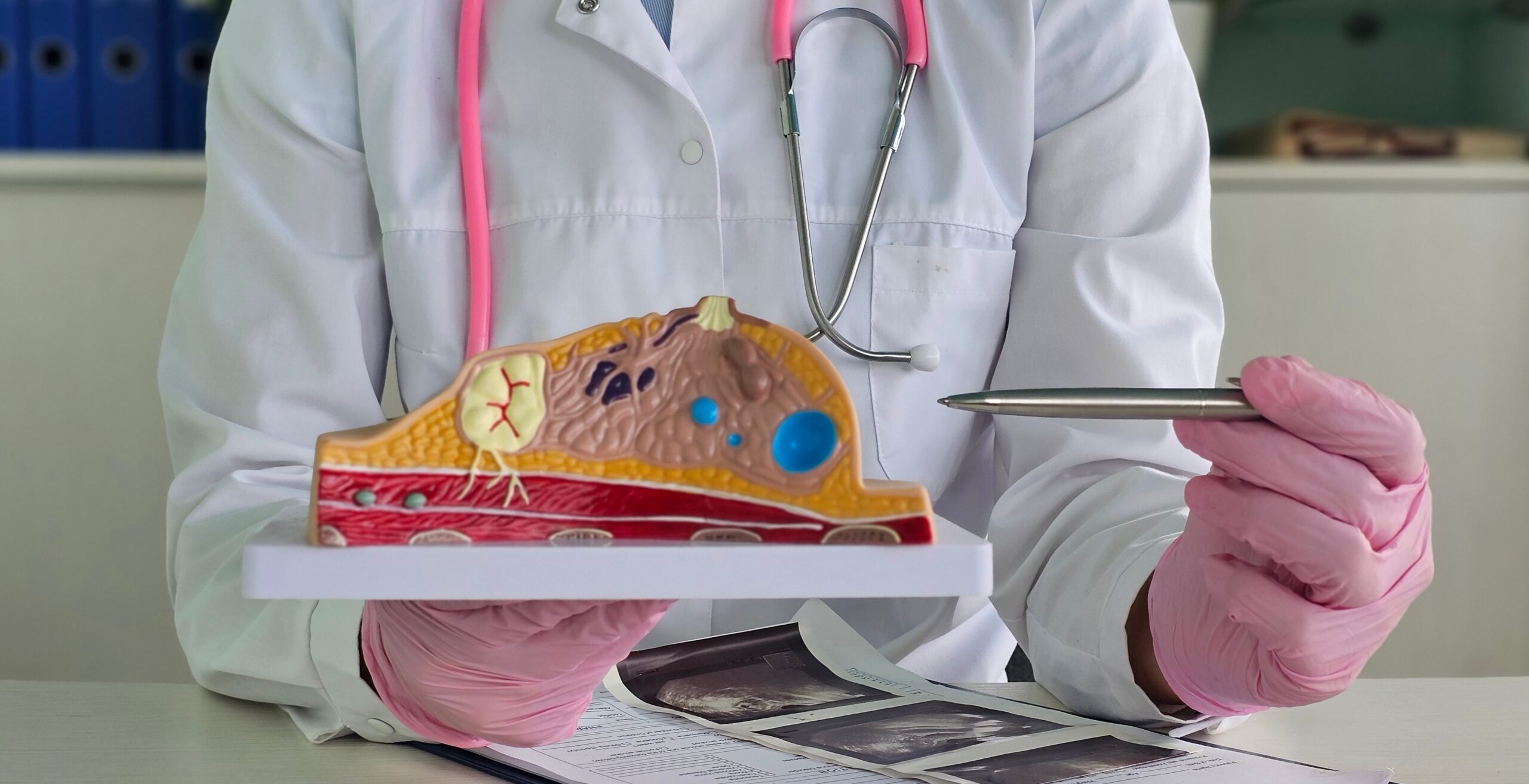
MYC Inhibitor Omomyc boosts PARP drug response in breast cancer
A preclinical study led by VHIO reveals that the MYC inhibitor Omomyc enhances the effectiveness of PARP inhibitors, offering a potential new treatment strategy for patients with drug-resistant triple-negative breast cancer.

A
Continue Reading
-
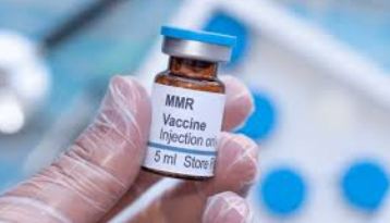
Doctor stresses urgent need for measles, rubella vaccination this winter

ISLAMABAD, Dec 5 (APP): A distinguished pediatrician Dr. Muhammad Aslam Friday underscored the urgent need for nationwide awareness regarding the government’s vaccination efforts, urging parents to…
Continue Reading
-

Nanotechnology Information | AZoNano.com – Page not found
Terms
While we only use edited and approved content for Azthena
answers, it may on occasions provide incorrect responses.
Please confirm any data provided with the related suppliers or
…Continue Reading
-

Targeting IL-23 and IL-17 in Psoriatic Disease: Pathways to Inflammation Control and PsA Prevention
In this episode, the panelists focus on the IL-23 and IL-17 pathways and their influence on inflammation control and psoriatic arthritis (PsA) prevention. The experts explain that IL-23 sits at the top of the inflammatory cascade, driving Th17…
Continue Reading
-
Doctor shares 3 natural ways to ease hot flashes for menopausal women – The Times of India
- Doctor shares 3 natural ways to ease hot flashes for menopausal women The Times of India
- Struggling with hot flashes? This new medication could help. Novant Health
- New drug reduced moderate to severe hot flashes by 92% Contemporary OB/GYN
- New…
Continue Reading