Prof Hamish McAllister-Williams, Professor of Affective Disorders, Newcastle University and Cumbria,…
Category: 6. Health
-
Human brains have five ‘eras’ across lifespan, scientists say – NewsNation
- Human brains have five ‘eras’ across lifespan, scientists say NewsNation
- Topological turning points across the human lifespan Nature
- This Is When We Actually Become Adults, According to Science Vogue
- So how old are you in your head? The…
Continue Reading
-

Overcoming disruption, transforming the AIDS response
The theme of this year’s World AIDS Day is “Overcoming disruption, transforming the AIDS response.”
The commemoration of World AIDS Day, which takes place on 1 December 2025, is an important opportunity to highlight the impact that the…
Continue Reading
-
Mirai outperforms Tyrer-Cuzick in breast cancer prediction – AuntMinnie
- Mirai outperforms Tyrer-Cuzick in breast cancer prediction AuntMinnie
- Matthew Kurian: AI in Breast Cancer Is Hitting a New Gear at SABCS25 Oncodaily
- Advancing mammography: evaluating the performance of artificial intelligence in estimating…
Continue Reading
-

How a high-carbohydrate diet increases risk of lung cancer: What to know, what to eat and what to avoid |
Many people reach for foods that provide comfort, convenience or quick energy, often without thinking about how rapidly those foods raise blood sugar. While high glycaemic index items are usually discussed in the context of weight gain or…
Continue Reading
-

Ethiopia health officials say prevention and control activities have been strengthened for Marburg virus
The Ministry of Health and the Ethiopian Public Health Institute (EPHI) reports through November 30, 12 confirmed Marburg Virus Disease (MVD) cases in the current outbreak, of which eight have died, as confirmed by the EPHI reference…
Continue Reading
-
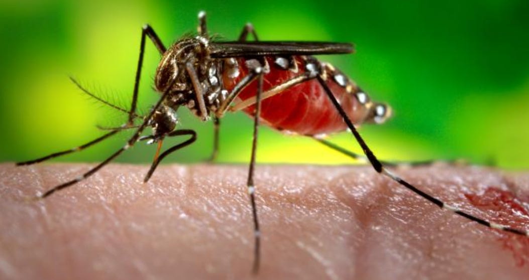
Vietnam reports 17% increase in dengue in 2025
Associate Professor, Dr. Nguyen Vu Trung – Director of the Pasteur Institute in Ho Chi Minh City said that the World Health Organization (WHO) ranked dengue fever as one of the 10 global health threats that need to be prioritized. Vietnam is…
Continue Reading
-

KRT17: A Key Driver of Cancer Therapy Resistance and Emerging Therapeu
Introduction
Cancer remains a leading cause of death worldwide, and the development of drug resistance poses a significant challenge to successful treatment, contributing substantially to cancer-related mortality. Therapy Resistance refers to…
Continue Reading
-

Ending HIV for mothers and children: a call to action
.
Trouble Chikoko, Country Director and Representative of UNAIDS; Pernille Ironside, UNICEF Representative in Pakistan; and Dr Luo Dapeng, WHO Country Representative in Pakistan
…Continue Reading
-
Scientists Reveal a Simple Eating Pattern That Helps Prevent Constipation – SciTechDaily
- Scientists Reveal a Simple Eating Pattern That Helps Prevent Constipation SciTechDaily
- Dietary Strategies to Enhance Regularity: Experts Advise on Combating Constipation with Fiber and Hydration The Boca Raton Tribune
- 7 Winter Fruits and…
Continue Reading