Commonly prescribed cardiovascular medications—such as statins, diuretics, and blood pressure drugs—appear to have little or no negative impact on survival among people living with multiple myeloma, according to new…
Category: 6. Health
-

Decoding the brain’s love for tempting snacks
Research from the University of East Anglia (UEA) may finally explain why we still reach for the biscuit tin, even when we’re full.
A new study reveals that the human brain continues to respond to tempting food cues even after we’ve…
Continue Reading
-

Living near nuclear power plants is associated with higher cancer mortality, national US study reports
A sweeping national analysis suggests that counties nearer to nuclear power plants experience higher cancer mortality, raising urgent questions about long-term environmental exposure, aging populations, and the limits of…
Continue Reading
-

Higher tyrosine levels linked to shorter lifespan in major UK Biobank analysis
A large genetic analysis suggests that one amino acid linked to protein metabolism could influence how long we live, with potential sex-specific effects that challenge assumptions about diet and longevity.
Study: The role of…
Continue Reading
-

What Is Salicornia? Health Benefits, Nutrition Facts, and Salt Substitute Potential
Introduction
What is Salicornia?
Micronutrients, minerals, and bioactives
The therapeutic potential of Salicornia
Sodium-related considerations
Culinary, industrial, and sustainable applications
Safety profile, research gaps, and toxicology
ReferencesContinue Reading
-
COLONTOWN Expands Patient Support With New Online Space for Colorectal Cancer Care
Nonprofit Colontown Provides Invaluable Resources and Support for Those With Colorectal Cancer and Their Carepartners:
“The Worldwide Online Community Recently Launched a New Platform
Feb. 9, 2026 – COLONTOWN, an online…
Continue Reading
-

shortcut or short-circuit to enhanced HIV outcomes?
aidsmap news story
At CROI 2026, scientists discussed how machine learning and generative artificial intelligence (AI) can be used to improve various HIV outcomes.
There are lingering, and often perplexing, questions as to exactly how these…
Continue Reading
-
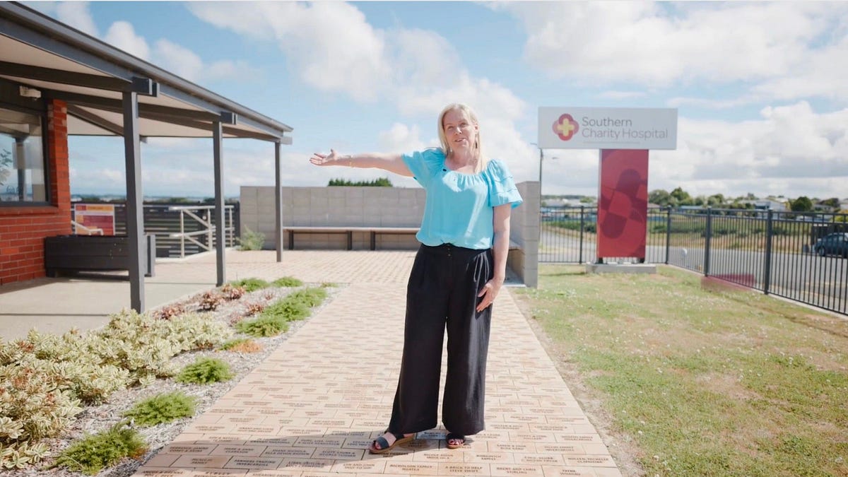
What the Southern Charity Hospital has achieved
Melissa Vining was the driver behind getting the Southern Charity Hospital opened on February 28, 2025. 
Saturday marked one year to the day that the ribbon was cut, and the Southern Charity Hospital opened.
It had taken a Herculean community effort…
Continue Reading
-

People amazed by woman’s tender dedication to her 48-year-old husband with dementia
In March 2023, after months of preparation and paperwork, Anita Omary arrived in the United States from her native Afghanistan to build a better life. Once she arrived in Connecticut, however, the experience was anything but easy.
“When I first…
Continue Reading
-

20-Year-Old Had Stomach Pain for Months, Diagnosed With Colon Cancer
Katie Davis lived the typical busy college student life when she first started feeling stomach pain.
Then a 20-year-old junior and marketing major at Westchester University in Pennsylvania, Davis split her time…
Continue Reading