- A cancer diagnosis can push people to crime The Economist
- The ‘Breaking Bad’ Effect From Cancer Is Real, Study Finds ScienceAlert
- New Study Links Cancer Diagnosis to Increased Crime Rates, Dubbed the ‘Breaking Bad Effect’ AOL.com
- Study Shows That…
Category: 6. Health
-
A cancer diagnosis can push people to crime – The Economist
-

Cancer After Cardiac Surgery: Long-Term Survival
CANCER after cardiac surgery was tied to shorter long-term survival, even though in-hospital mortality remained low.
Cancer After Cardiac Surgery and Long-Term Survival
As more patients present for cardiac surgery with active cancer or a prior…
Continue Reading
-

Doing An Annual Mental Health Check-Up Via The Use Of AI Chatbots Such As ChatGPT
Should people be encouraged to undertake an annual mental health check-up via AI or is that a bridge too far?
getty
In today’s column, I examine a somewhat novel idea that people should consider using generative AI and large language models as a…
Continue Reading
-
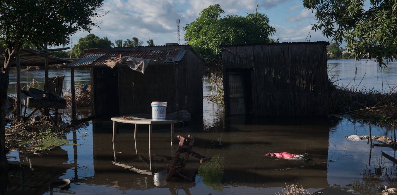
how disaster systems can prepare better
When floods sweep through southern Africa, the most visible damage is immediate: homes washed away, crops destroyed, clinics disrupted, families displaced. These images dominate headlines and humanitarian appeals.
But as floodwaters recede,…
Continue Reading
-
The impact of different exercise modes on prostate cancer: a Bayesian network meta-analysis
Schafer, E. J. et al. Recent Patterns and Trends in Global Prostate Cancer Incidence and Mortality. Update Eur. Urol. 87 (3), 302–313 (2025).
Van Blarigan, E. L. et al. Trends in Prostate…
Continue Reading
-

‘We’re seeing ‘SuperAgers’ aged 80+ with memory as sharp as 40-year-olds — neurogenesis powers this’
Please describe your research?■ The SuperAgers study at Northwestern University was developed 25 years ago. Its goal is to examine individuals over the age of 80 who have the memory capacity of people aged 30 or 40 years younger. This aims to…
Continue Reading
-
Measles cases increase across the Southwestern Utah region as more people opt out of MMR vaccines – St. George News
- Measles cases increase across the Southwestern Utah region as more people opt out of MMR vaccines St. George News
- Me, Myself, as Mommy: Utah’s measles outbreak highlights frustrating times standard.net
- Measles declared community spread as six…
Continue Reading
-

Does When You Eat Really Matter
Men’s Fitness aims to feature only the best products and services. If you buy something via one of our links, we may earn a commission.
Meal timing has become nutrition’s favorite battlefield. Some swear by fasting windows, others by eating…
Continue Reading
-
Nearly half of Americans unaware processed meat is tied to colorectal cancer, poll finds – Medical Xpress
- Nearly half of Americans unaware processed meat is tied to colorectal cancer, poll finds Medical Xpress
- New Poll: Almost Half of US Adults Unaware of Connection Between Processed Meat Consumption and Colorectal Cancer Physicians Committee for…
Continue Reading
-

Explainer | How TCM’s ‘miracle pill’ is used to treat strokes, but experts urge caution
This series is based on our reporting on TCM: its history, treatments and growing acceptance around the world. This is the seventh instalment.
In the world of traditional Chinese medicine (TCM), few remedies carry the legendary status – or the…
Continue Reading