
How well do students at your school understand what bullying is? Do they feel comfortable intervening when they witness bullying behaviour on the school grounds? Does your…
Category: 6. Health
-

Long ADHD wait times leave families feeling powerless and ‘forever in limbo’
Families are feeling stressed, powerless and ‘forever in limbo’ as they wait months, or sometimes years, for Attention-Deficit Hyperactivity Disorder (ADHD) assessments, according to a new study.
Research led by the…
Continue Reading
-

Six Immune Cell Phenotypes Implicated in Clear Cell Ovarian Cancer Origins
Worldwide ovarian cancer ranks fourth among disease-related causes of death in women with 240,000 new cases each year. Prognosis varies significantly by stage at diagnosis and histologic type. Clear cell ovarian cancer (CCOC), a specific subtype…
Continue Reading
-

Healthy thymus gland linked to longer life and immune stability
People with a healthy thymus gland live longer and are less likely to fall ill. In addition, immunotherapies are more often successful in patients with a healthy thymus. This is shown by two international studies involving…
Continue Reading
-

US Withdrawal Of Global Health Funding Is ‘Public Health Emergency Of International Concern’
US President Donald Trump shocked partners when he announced an immediate freezing of all international health aid shortly after assuming power in January 2025. The rapid withdrawal of international health aid by the United States (US)…
Continue Reading
-
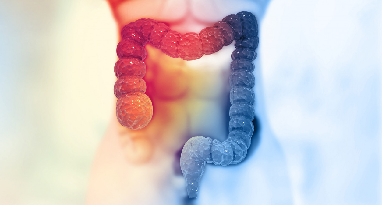
Chronic Inflammation Leaves Long-Lasting Impression on Gut Stem Cells, Increasing Colorectal Cancer Risk
In a new study, funded in part by the National Institutes of Health (NIH), researchers have uncovered a molecular mechanism that could explain how chronic gut inflammation may increase the risk of colorectal cancer. By…
Continue Reading
-

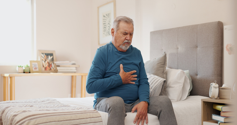
Benefits of Early SAVR for Asymptomatic Aortic Stenosis Persist a Decade On
The final results of RECOVERY add support to addressing severe aortic stenosis right away rather than waiting for symptoms.
Patients with asymptomatic severe aortic stenosis (AS) who undergo immediate SAVR have better outcomes than those…
Continue Reading
-
Study Finds No Negative Impact of COPD Treatment With Vitamin K Antagonists
The use of vitamin K antagonists (VKAs) by patients with COPD and atrial fibrillation is not associated with worse respiratory outcomes, according to findings of a national cohort study by Bård-Emil Vang Vang Gundersen, MD, and…
Continue Reading
-

Scientists Find Surprising Combination That Boosts Weight Loss After Menopause
Weight gain is common in menopause, with some data suggesting that women gain an average of a pound a year during this life stage. If you’re in the throes of it, about to go through it, or are on the other side, it’s fair to be curious about…
Continue Reading
-
Scientists discover “overflow valve” in cells linked to Parkinson’s Disease
Researchers have uncovered how a mysterious ion channel helps cells break down waste, opening new possibilities for treating Parkinson’s disease.
Just like sinks and bathtubs have overflow drains to prevent spills, human cells appear to have a…
Continue Reading