Pregnant women with opioid use disorder who were prescribed buprenorphine had fewer emergency department visits and hospital admissions, with the greatest benefit seen among those who adhered to treatment on at least 80% of days.
Continue Reading


A nurse-led healthy lifestyle group intervention enhanced supportive care for patients living with metastatic breast cancer, according to the findings of a small, open pilot study.
Led by Loren N. Winters, ANP-BC, OCN, Dipl ABLM, CHWC, Mass…
The Ministry of Health reiterates the effectiveness of community participation in reducing the spread of diseases.
The Ministry of Health, through the Department of Epidemiology, reported the figures corresponding to epidemiological week No. 42,…

Medical Express In patients with long COVID, a new study has revealed a structural association between circulating microclots and neutrophil extracellular traps (NETs). This finding suggests the…
Washington Post After the Centers for Disease Control and Prevention shifted its coronavirus vaccine guidance from a near-universal recommendation to a more limited approach, many readers asked…
This request seems a bit unusual, so we need to confirm that you’re human. Please press and hold the button until it turns completely green. Thank you for your cooperation!
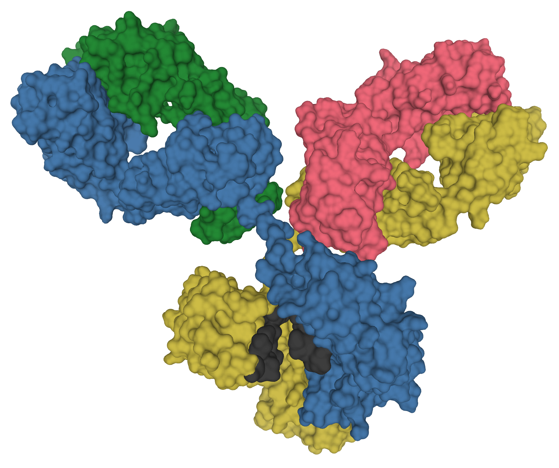
An antibody from a Tanzanian woman discovered during screening for anti-HIV antibodies shows a strong therapeutic potential in a preclinical study. Named 04_A06, it neutralised (blocked) 97.3% of over 300 HIV strains tested, and blocked 77% of…
This request seems a bit unusual, so we need to confirm that you’re human. Please press and hold the button until it turns completely green. Thank you for your cooperation!
Reposted from U of U Health.
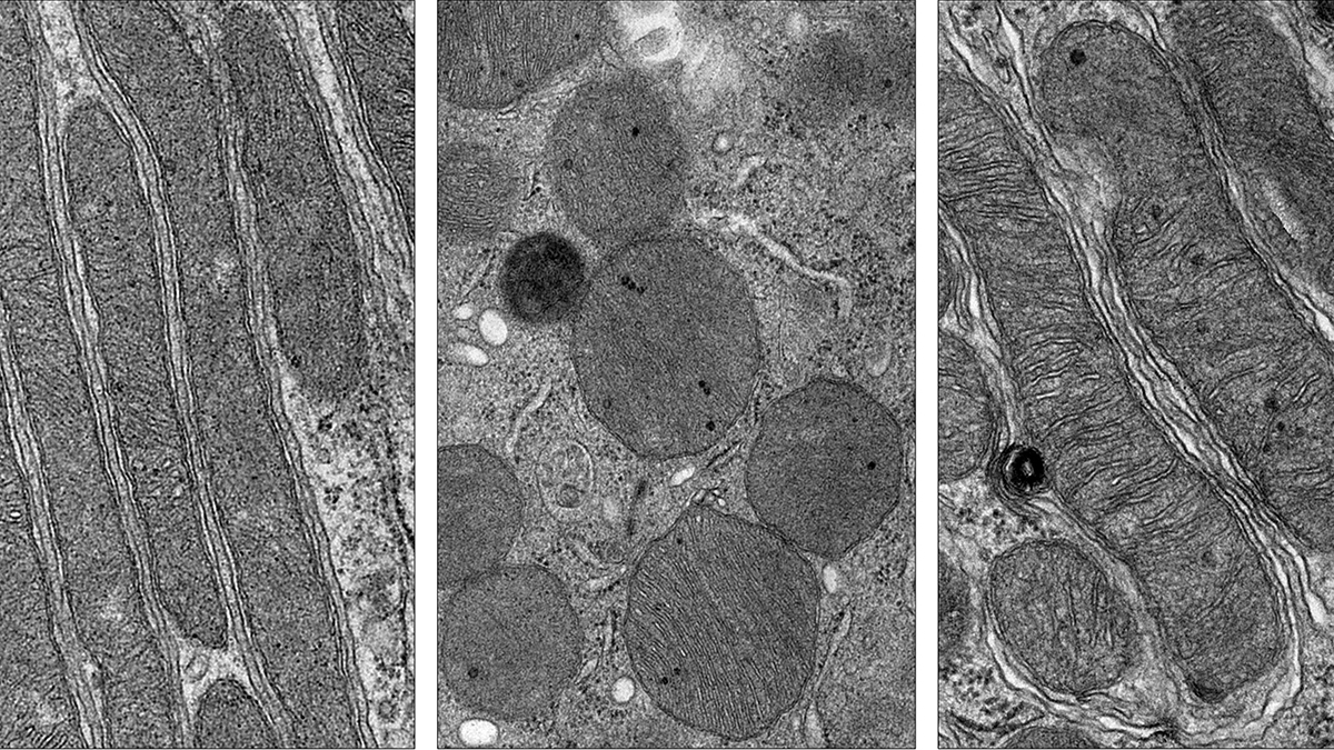
Serious damage to short-term kidney function—known as acute kidney injury, or AKI—can be fatal and also increase the risk of irreversible chronic kidney disease. It can be triggered by stressors…
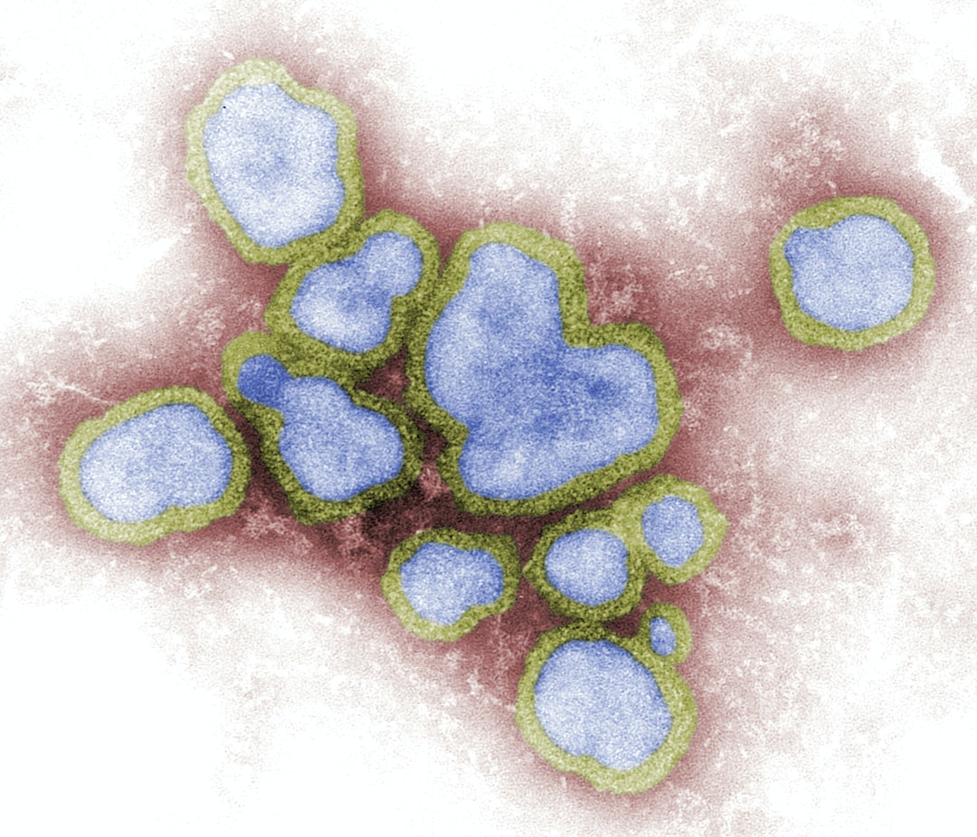
The Conversation Since first being detected in Newfoundland in 2021, a subtype of highly pathogenic avian influenza, HPAI A(H5Nx), has had a dramatic impact on North America.
The poultry industry has suffered the most, with almost 15…