Researchers have long established that hormones significantly affect the brain, creating changes in emotion, energy levels, and decision-making. However, the intricacies of these processes are not well understood.
A new study by a…
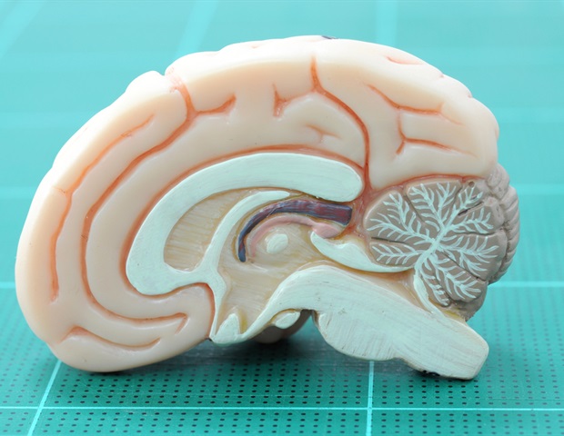
Researchers have long established that hormones significantly affect the brain, creating changes in emotion, energy levels, and decision-making. However, the intricacies of these processes are not well understood.
A new study by a…

Metabolic dysfunction-associated steatotic liver disease (MASLD) represents an escalating healthcare burden across the Middle East and North Africa (MENA) region; however, system-level preparedness remains…

Anxiety disorders are some of the most common mental health conditions in America, affecting about one in five people nationwide. But much remains unknown about the roots of anxiety in the brain. Now, research at the University of…

Menopause stage doesn’t impact brain volume; age does, according to research presented at

I’ve always had very fair skin and light-colored eyes. Over the years, I’ve had plenty of sun exposure, too — first as a geophysicist working outdoors and later as a swim mom spending hours on the pool deck. I knew I was at higher risk for…

Newswise — PHILADELPHIA — (Nov. 11, 2025) — On Monday, Nov. 17th at 6:30 pm EST, Richard Jefferys, Basic Science, Vaccines and Cure Project director at the Treatment Action Group (TAG), delivers the 29th annual…

BYLINE: Dacia Morris
Newswise — New York, NY – Nov. 11, 2025 – Pneumonia, a largely preventable disease, caused more than two million deaths in 2021, according to the…

New research from Edith Cowan University (ECU) has revealed a striking disconnect between how recreational athletes perceive their health and fitness, and how they feel about their bodies.
The research found that while 69 per…