A new genomic method has enabled multiple people with rare conditions to receive diagnoses that were previously unattainable by identifying complex structural genetic changes that are often missed by standard tests.
Researchers at…

A new genomic method has enabled multiple people with rare conditions to receive diagnoses that were previously unattainable by identifying complex structural genetic changes that are often missed by standard tests.
Researchers at…

A deep learning algorithm developed for processing digital screening mammograms, Mirai, was able to detect interval breast cancers and identify women who would benefit from more frequent screenings, according to the results of a UK…
Backed by real-world evidence from more than 500 studies, a new review shows that this season’s Covid-19, RSV, and flu vaccines continue to protect against severe illness.
Study: Updated Evidence for Covid-19, RSV, and Influenza…

More than 4 in 5 women are gritting out menopause without seeking any treatment for their symptoms, researchers said in a new study released by the Mayo Clinic. Photo by Adobe Stock/HealthDay News
Women are gritting out menopause without seeking…
For someone who has heart surgery or experiences a major cardiovascular event, cardiac rehabilitation can significantly improve their short and long term recovery.
People who participate in this comprehensive exercise, lifestyle…

As mpox continues to affect communities across 26 African countries, health officials are turning to familiar strategies to manage the caseload. One such approach, home-based care, is being adapted from the COVID-19 response, offering a…
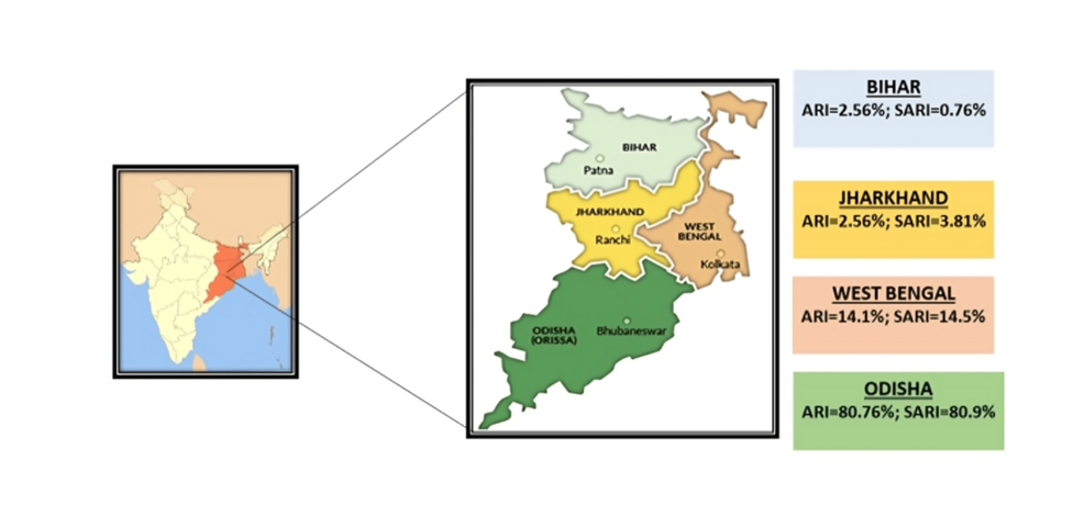
Clostridioides difficile (C. difficile) is a leading cause of hospital-acquired diarrhea worldwide. The incidence and mortality of Clostridioides difficile infection (CDI) have risen significantly in recent years, posing a…

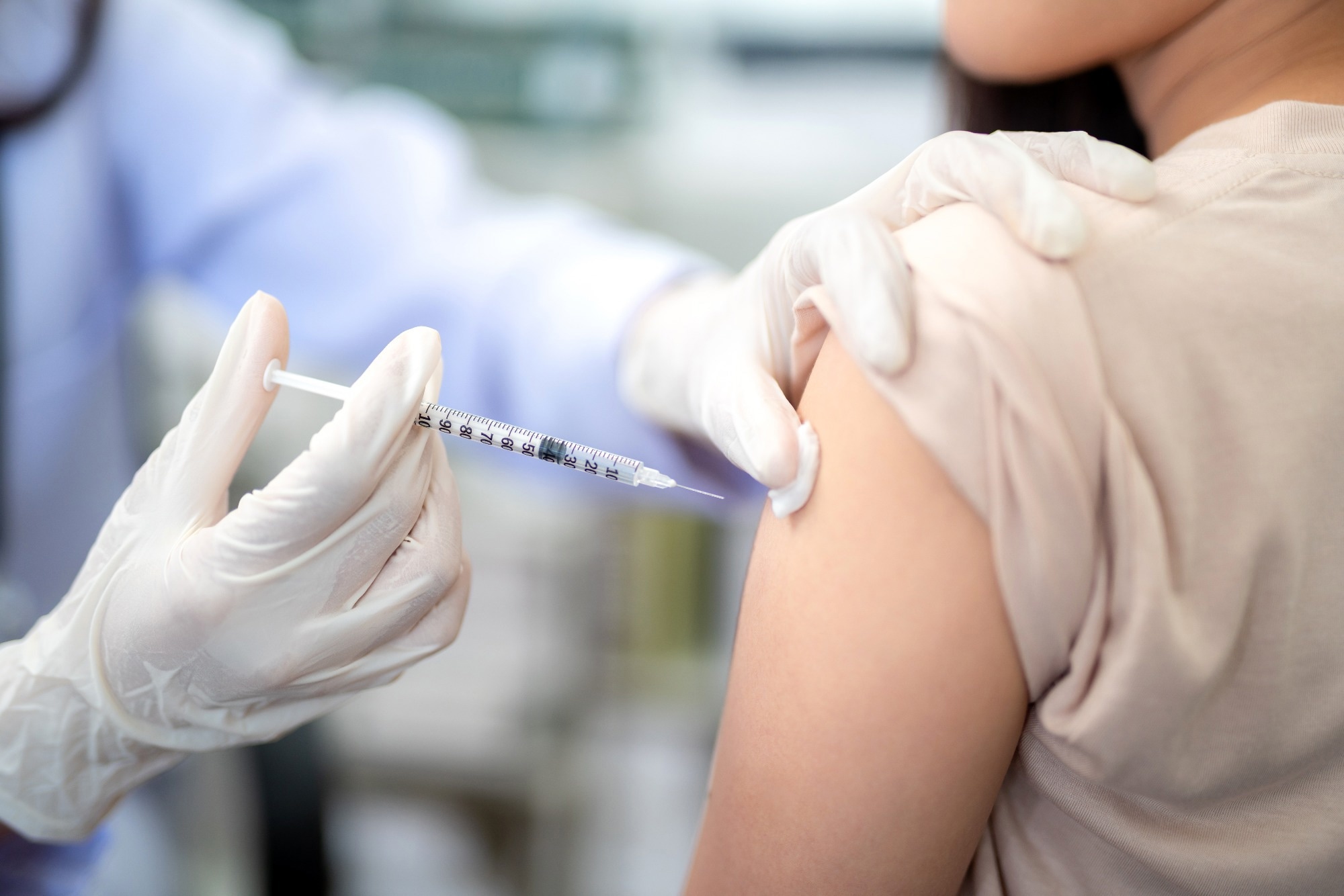
Researchers may have developed the first vaccine against Salmonella enterica serovar Paratyphi A (S. Paratyphi A). In a phase 2b randomized trial (ISRCTN15485902), 2 doses of the oral, investigational live attenuated vaccine CVD 1902 showed 73%…
This request seems a bit unusual, so we need to confirm that you’re human. Please press and hold the button until it turns completely green. Thank you for your cooperation!