Researchers developed a deep neural network, M-PACT, to identify and classify brain tumors in pediatric patients from the subnanogram-input cell-free DNA of methylomes, according to findings published in Nature Cancer.
“This is a…

Researchers developed a deep neural network, M-PACT, to identify and classify brain tumors in pediatric patients from the subnanogram-input cell-free DNA of methylomes, according to findings published in Nature Cancer.
“This is a…

Researchers at UC San Francisco have identified a biological process that may explain why exercise sharpens thinking and memory. Their findings suggest that physical activity strengthens the brain’s built in defense system, helping protect it…

Weetabix, the UK’s leading cereal manufacturer, has announced a massive packaging refresh across its core portfolio, aimed at addressing widespread nutritional deficiencies in the British public.
Moving beyond traditional branding, the new…
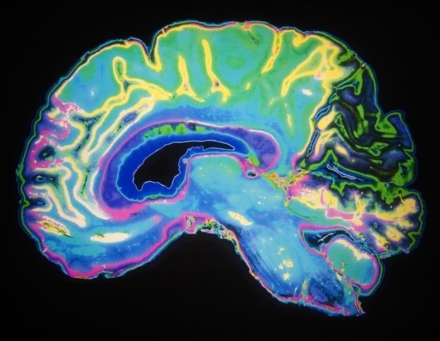
An international research team, with significant involvement from the Medical University of Vienna, has developed a new AI-based analysis method that can accurately classify brain tumors using genetic material from cerebrospinal…

A large, diverse cohort study shows that muscle strength, especially grip strength, may signal survival odds in older women, regardless of how much they move, how long they sit, or their measured fitness level.
Study: Muscular…

A Canadian study has linked binge drinking at age 50 and above to an increased mortality risk of 23% compared to the participants who did not binge drink in the year before.
The study followed 129,000 adults aged 50 and above for up to 12 years…

Parkinson’s disease is a long term neurological condition that gradually worsens over time. More than one million people in the United States are living with the disorder, and about 90,000 new cases are diagnosed each year. Current medications…
Acinetobacter baumannii is a conditionally pathogenic gram-negative bacterium that has emerged as a major pathogen causing hospital infection outbreaks because of its…

Older adults who live with higher levels of air pollution are more likely to develop Alzheimer’s disease, according to new research led by Yanling Deng of Emory University, U.S.A. The study was published February 17th in the open access journal…