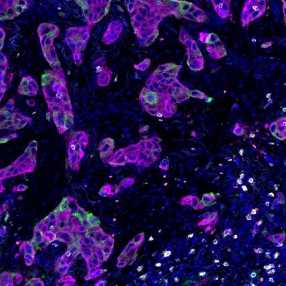
Category: 8. Health

  • The Diagnosis and Prognosis Value of Exosomal MascRNA in Patients with

    The Diagnosis and Prognosis Value of Exosomal MascRNA in Patients with

    Introduction

    Cardiovascular disease (CVD) represents the leading cause of mortality and morbidity globally, accounting for approximately 30% of all disease-related deaths each year.1 Acute coronary syndrome (ACS) is one of the most lethal subtypes of coronary heart disease, requiring timely risk assessment and effective therapeutic interventions to improve patient outcomes.2 Advances in medical techniques, particularly the widespread use of percutaneous coronary intervention (PCI), have significantly reduced mortality of ACS.3,4 However, the major adverse cardiovascular events (MACEs) after PCI continue to threaten the health and quality of life of ACS patients.5,6 Therefore, identifying biomarkers that can improve early diagnosis and predict the prognosis of ACS remains an urgent priority.7

    Exosomes are small extracellular vesicles, with dimensions ranging from 30 to 150 nm, secreted by nearly all cell types. They function as cargo transporters, transferring nucleic acid, proteins, lipids, and other stuffs between cells.8 Exosomes are integral to numerous biological processes and the pathogenesis of various diseases.9 The process of exosome biogenesis enables the packaging of molecules from both membranous and cytosolic origins, making them reflective of the state of the releasing cell and providing valuable insights into the cellular environment. The encapsulation of proteins and RNAs within exosomes prevents their degradation, making exosomes an ideal source of biomarkers. Advances in exosome isolation techniques have garnered significant attention for their potential in clinical applications. Increasing evidence supports the potential of exosomes as valuable biomarkers for early diagnosis and prognosis assessment in cardiovascular diseases.10

    MALAT1-associated small cytoplasmic RNA (mascRNA) originates from the nuclear long non-coding RNA MALAT1, is a tRNA-like small non-coding RNA, and is localized in the cytoplasm.11 While MALAT1 has been extensively studied and shown to influence various cellular processes, including the development of atherosclerosis,12,13 the function of mascRNA remains largely unknown. Recent research has detected high levels of mascRNA in circulating human peripheral blood mononuclear cells (PBMCs).14 MascRNA suppresses the production of inflammatory cytokines in LPS-stimulated macrophages by inhibiting the activation of NF-κB and MAPK signaling pathways.15 In murine models of atherosclerosis, mascRNA deficiency leads to hyperactivity of circulating inflammatory cells and an increased macrophage presence in atherosclerotic plaques, contributing to plaque rupture and thrombosis formation.16 However, the expression of mascRNA in circulating exosomes remains poorly understood.

    Our previous study demonstrated that MALAT1 may serve as a promising biomarker for cardiovascular disease, showing diagnostic potential for ACS patients.17 However, limited research has evaluated the diagnostic value of mascRNA in cardiovascular disease. This study aims to explore the link between plasma-derived exosomal mascRNA and the occurrence of ACS and its association with adverse cardiovascular events.

    Materials and Methods

    Study Subjects

    This study included 281 patients who underwent coronary angiography at Meizhou People’s Hospital from Oct. 2021 to May 2024. The ACS patients were diagnosed according to the 2020 ESC Guidelines for managing acute coronary syndromes,18 which were characterized by symptoms such as recurrent chest pain at rest or with minimal exertion, as well as severe angina that began or worsened within 4 weeks before the procedure. The exclusion criteria included severe valvular heart disease, severe arrhythmias, acute or chronic inflammation, malignant tumors, autoimmune diseases, and hematologic disorders. The non-ACS group included individuals who were diagnosed without coronary artery disease (CAD) by cardiologists as coronary angiography indicative of stenosis < 50%. This research was approved by the Ethical Committee of Meizhou People’s Hospital (Approval No. MPH-HEC 2023-C-34) and was conducted in full accordance with the principles of the Declaration of Helsinki. Informed written consent was collected from each participant. Figure 1 illustrates the study flow.

    Figure 1 Study flow diagram.

    Plasma Collection

    A total of 5 mL of venous blood was obtained from patients prior to PCI and placed in EDTA anticoagulant tubes. The samples were maintained at 4°C for 2 hours. Subsequently, the samples were centrifuged at 300g for 10 minutes, after which the supernatant was collected and aliquoted into centrifuge tubes for exosome isolation.

    Exosome Isolation

    Exosomes were isolated from plasma by ultracentrifugation techniques (CP100NC, Hitachi), as shown in Figure 2A. In brief, the plasma was subjected to centrifugation at 2000g for 10 minutes, after which the supernatant was collected. This was followed by centrifugation at 10,000g for 30 minutes, and the supernatant was again collected. Subsequently, the sample was centrifuged at 120,000g for 30 minutes, and the supernatant was carefully discarded. The resulting pellet was resuspended in PBS and centrifuged once more at 120,000g for 30 minutes. The supernatant was discarded, leaving the exosomes at the bottom for subsequent experiments. All centrifugation procedures were performed at 4°C.

    Figure 2 Isolation and characterization of exosomes from plasma. (A) Centrifugation protocol for enrichment of plasma exosomes; (B) Transmission electron microscopy (TEM) analysis of the exosome morphology. Representative exosomes are indicated by arrows; (C) The particle size of exosomes was measured by nanoparticle tracking analysis. (D) Western blot analysis of the exosomal markers.

    Characterization of Exosomes

    The exosomes derived from plasma were characterized using nanoparticle tracking analysis (NTA), Western blotting, and transmission electron microscopy (TEM). For the Western blot analysis, the exosomes were probed with the following primary antibodies: anti-CD9 (1:1000, Cell Signaling Technology), anti-CD63 (1:1000, Cell Signaling Technology), and anti-TSG101 (1:1000, Cell Signaling Technology). The NTA was conducted using a NanoSight NS300 instrument (Malvern Panalytical) to evaluate the size, distribution, and concentration of the exosomes. TEM was performed with a JEM-1400 microscope (JEOL, Japan) to examine the ultrastructural features and size of the exosomes.

    RNA Isolation and Reverse Transcription-Quantitative Polymerase Chain Reaction (qRT-PCR)

    Exosomal RNA was extracted utilizing the SteadyPure Small RNA Extraction Kit (Accurate Biology, China). RNA quality was assessed by measuring the A260/A280 ratio with an ultramicro-spectrophotometer (NP80, IMPLEN, Germany). Complementary DNA (cDNA) was generated by PrimeScript™ RT reagent Kit (Takara, Japan). Exosomal mascRNA expression was determined utilizing the TB Green® Premix Ex Taq™ II and normalized to U6 using the 2−ΔΔCt method.19 The primer sequences for qRT-PCR were as follows:

    mascRNA forward, 5’-GATGCTGGTGGTTGGCACTC-3’; mascRNA reverse, 5’-TGGAGACGCCGCAGGGAT-3’; U6 forward, 5’-CTCGCTTCGGCAGCACA-3’; U6 reverse, 5’-AACGCTTCACGAATTTGCGT-3’.

    Clinical Data Collection and Follow-Up

    The clinical characteristics of patients were retrieved from the hospital’s electronic medical records. Collected variables included age, gender, hypertension, diabetes mellitus, dyslipidemia, left ventricular ejection fraction (LVEF), blood pressure, glucose levels, lipid profiles and blood cell counts.

    One-year follow-up data for ACS patients were obtained from electronic medical records or through telephone interviews. The primary outcome measure was the incidence of major adverse cardiovascular events (MACE) including all-cause mortality, nonfatal myocardial infarction, target vessel revascularization, rehospitalization for angina or heart failure, and stent thrombosis.

    Statistical Analysis

    Statistical analyses were conducted using SPSS 20.0 (IBM Corp., Armonk, NY, USA). Data were presented as mean ± SD or number (percentage). The Shapiro–Wilk test checked the normality of continuous variables. Student’s t-test was used for continuous variables, and chi-square or Fisher’s exact test for categorical variables. The sample size, based on China’s ACS incidence of 1%, is approximately 95, with a significance level of α = 0.05 and a 2% margin of error. The correlation between exosomal mascRNA and clinical parameters were analyzed by Spearman correlation analysis. Logistic multivariate regression analysis was employed to assess the relationship between exosomal mascRNA and ACS risk. Receiver operating characteristic (ROC) curve analysis was employed to evaluate the diagnostic value of exosomal mascRNA for ACS. The one-year MACE-free survival was assessed using Kaplan–Meier analysis and the Log rank test, while multivariable Cox regression identified predictors of 1-year MACEs in ACS patients. A P-value < 0.05 was considered statistically significant.

    Result

    Characteristics of Study Subjects

    The study included 140 ACS patients and 141 non-ACS, with baseline characteristics summarized in Table 1. There was no difference between the two groups regarding gender, age, hypertension, diabetes mellitus, and dyslipidemia. ACS patients exhibited higher levels of white blood cell (WBC), monocytes, neutrophils, Gensini scores, cTnI (P < 0.05), and lower LVEF compared to non-ACS group (P < 0.05).

    Table 1 Baseline Characteristics of Study Subjects

    Identification of Plasma Exosomes

    Plasma exosomes were isolated utilizing multiple ultracentrifugation steps (Figure 2A). Plasma exosomes exhibited a typical double-layered vesicular structure (Figure 2B), with a mean diameter of approximately 130 nm (Figure 2C). Western blot analysis verified the expression of exosomal protein markers CD9, TSG101 and CD63 (Figure 2D).

    Expression of Exosomal mascRNA in Patients with ACS

    Our data suggested that exosomal mascRNA expression was elevated in ACS patients compared to the non-ACS (Figure 3A). However, exosomal mascRNA expression showed no significance between the subgroups of ACS (Figure 3B).

    Figure 3 The expression of exosomal mascRNA in patients with ACS. (A) Exosomal mascRNA levels in ACS patients and non-ACS patients. **P < 0.01, comparison was tested by Student’s t test; (B) Exosomal mascRNA levels in different types of ACS patients. (C) Comparison of exosomal mascRNA expression in ACS patients with MACE and non-MACE during the one-year follow-up. *P < 0.05, comparison was tested by Student’s t test.

    We compared the expression of exosomal mascRNA in patients with or without MACEs during the 1-year follow-up period after PCI treatment. A total of 29 ACS patients developed MACEs during the follow-up. Our data showed that mascRNA expression was significantly higher in the MACE group than the non-MACE group (Figure 3C).

    Association Between Exosomal mascRNA and Clinical Variables

    We further analyzed the association between exosomal mascRNA and clinical parameters. As shown in Figure 4, the Spearman correlation analysis revealed a significant positive correlation between exosomal mascRNA levels and Gensini scores (r = 0.242, P < 0.001), LDL (r = 0.173, P = 0.019), WBC (r = 0.183, P = 0.012), age (r = 0.164, P = 0.013). No significant associations were observed between exosomal mascRNA levels and LVEF (r = −0.120, P = 0.103), neutrophil count (r = 0.100, P = 0.109), as these differences did not reach statistical significance.

    Figure 4 Correlation between exosomal mascRNA and clinical parameters. The correlation between exosomal mascRNA and age (A), LVEF (B), Gensini score (C), LDL level (D), WBC (E), and neutrophil (F) was assessed by Spearman correlation analysis.

    The Diagnostic Value of Exosomal mascRNA for ACS

    The diagnostic value of exosomal mascRNA for ACS was evaluated by ROC curve analysis. Our data revealed that exosomal mascRNA serves as a diagnostic predictor for ACS, with an AUC of 0.763 (95% CI: 0.702–0.824) and cutoff value of 1.173 (Figure 5). The predictive performance of mascRNA improved when combined with cTnI, with the AUCs increased to 0.866 (95% CI: 0.815–0.916) (Figure 5).

    Figure 5 The diagnostic value of exosomal mascRNA for ACS.

    To illustrate the association of the exosomal mascRNA with ACS risk, its levels were categorized into quartiles (35 patients for each quartiles). Compared with patients in the first quartile for mascRNA expression, patients in the second, third and fourth quartiles exhibited increased ACS risk (OR: 3.423, 95% CI: 1.427–8.213, OR: 5.542, 95% CI: 1.859–16.524 and OR: 9.288, 95% CI: 3.275–26.340, respectively; all P < 0.01; Table 2).

    Table 2 Association Between Exosomal mascRNA Expression and Risk of ACS

    The Prognostic Value of Exosomal mascRNA for ACS

    We further explored whether the expression of exosomal mascRNA predict the occurrence of MACEs. Patients were divided into high mascRNA group (≥ 3.85, n = 60) and low mascRNA group (< 3.85, n = 60). Kaplan-Meier analysis and Log rank test were utilized to assess the 1-year MACEs‐free survival rate between high mascRNA and low mascRNA groups. The data revealed that patients with high mascRNA expression have a lower incidence of MACE-free survival compared to those with low mascRNA expression (long rank P < 0.001) (Figure 6).

    Figure 6 The prognostic value of exosomal mascRNA in patients ACS. The 1-year MACEs‐free survival rate between high mascRNA and low mascRNA groups was assessed by Kaplan-Meier curves.

    A multivariate Cox regression analysis was performed to determine association between exosomal mascRNA and MACEs in ACS patients. After adjusted for age, diabetes mellitus and LVEF, mascRNA was significantly associated with the occurrence of 1-year MACEs, with a HR of 2.959 (95% CI: 1.187–4.669, P < 0.001) (Tables 3 and 4).

    Table 3 Clinical Characteristics of Non-MACE and MACE Group in ACS Individuals

    Table 4 Multivariate Cox Regression Model Analysis of MACEs in ACS Patients

    Discussion

    ACS is still the leading cause of mortality despite the advances in treatment and diagnostic modalities.2 Precise diagnosis of ACS is crucial for effective therapeutic intervention and enhancing patient survival rates. The study found that exosomal mascRNA levels were significantly higher in ACS patients and closely linked to ACS risk, suggesting its potential as a diagnostic and prognostic biomarker.

    Cardiac troponin (cTnI) is the key plasma biomarker for detecting myocardial injury, with high sensitivity and specificity for diagnosing acute myocardial infarction (AMI). However, its specificity is low in the first 3 hours after symptoms begin, and elevated levels can also indicate other conditions such as myocarditis and stress-induced cardiomyopathy.20,21 Exosomes have attracted increasing interest in the cardiovascular field due to their potential clinical implications. More and more exosome-based biomarkers are identified for diagnosis of cardiovascular diseases.22–25 This study found that exosomal mascRNA levels were significantly higher in ACS patients, regardless of the type of ACS (UA, STEMI, or NTEMI), and were linked to an increased risk of ACS. MascRNA levels correlated with the Gensini score, LDL, and WBC, which are related to vascular stenosis, inflammation, and lipid metabolism. Notably, although the findings are significant, the correlations are weak, thus more studies would be needed to validate the clinical relevance of mascRNA.

    MascRNA is a highly conserved small non-coding RNA originating from the primary transcript of MALAT1.11 As one of the most abundant lncRNAs, MALAT1 has been established as a crucial regulator in cardiovascular pathological processes.26–28 Our previous study as well as studies of others suggested that MALAT1 was enriched in exosomes and serve as potential biomarker for coronary heart disease.29,30 To the best of our knowledge, this study is the first to identify the expression of mascRNA in plasma exosomes. Our data suggested that exosomal mascRNA could distinguish ACS from non-ACS individuals, achieving an AUC of 0.776. Notably, the combination of mascRNA and cTnI markedly enhanced diagnostic performance, achieving an AUC of 0.884, surpassing the efficacy of either marker alone and underscoring its clinical utility. Further investigation is warranted to assess the optimal integration of mascRNA with other established or emerging biomarkers to improve the specificity and accuracy of ACS diagnosis.

    The prediction of major adverse cardiovascular events (MACE) is crucial for optimizing treatment strategies in patients with acute coronary syndrome (ACS). Numerous inflammatory biomarkers, such as C-reactive protein (CRP), the neutrophil-lymphocyte ratio (NLR), the fibrinogen/albumin ratio (FAR), and the systemic immune-inflammation index (SII), are gaining prominence in research due to their cost-effectiveness, simplicity, and ease of application.31–33 Although these inflammatory biomarkers demonstrated a strong correlation with the occurrence of major adverse cardiovascular events (MACEs), their specificity remains problematic. Consequently, predictive biomarkers for MACEs are still limited.34 This study found that patients with high exosomal mascRNA levels experienced a higher rate of MACEs within a year after PCI treatment, with mascRNA being an independent risk factor (HR = 3.357). This suggests a link between mascRNA and ACS outcomes. While the typical MACE incidence post-PCI is around 10%, our one-year follow-up showed a 20.7% rate (29/140), possibly due to the MACE criteria and the predominance of AMI among ACS patients.

    Although the exact mechanisms underlying how mascRNA participated in the pathology of ACS remained unclear, some research suggested that it is in part due to its function on inflammation. Sun et al35 reported that mascRNA inhibits the activation of NF-κB and MAPK signaling, as well as the production of inflammatory cytokines in macrophages stimulated by LPS. Gast et al16 found that selective ablation of mascRNA resulted in massive induction of TNF and IL-6 in macrophages, which significantly exacerbated vascular injury compared to wildtype macrophages. Previous studies have shown that endothelial dysfunction is linked to future MACEs. Endothelial dysfunction is a key factor in myocardial infarction and central to all ACS, contributing to atherosclerosis through vasoconstriction, macrophage migration, cellular growth, and inflammation.36–38 Our prior research indicated that MALAT1 inhibits endothelial inflammation and the interactions between monocytes and endothelial cells through ATG5-mediated autophagy.13 Since mascRNA is closely associated with MALAT1, mascRNA may also participate in the regulation of endothelial inflammation. Nonetheless, additional investigations are required to elucidate the underlying mechanisms.

    This study is subject to several limitations. Firstly, as a single-center investigation with a relatively small sample size and a retrospective design, it is vulnerable to information and selection biases. Consequently, multicenter cohort studies are required to validate our findings. Secondly, this study did not include a comparison of mascRNA levels before and after patient treatment. Future research should assess the changes in mascRNA expression pre- and post-treatment to explore its predictive value for MACEs.

    Conclusions

    In summary, exosomal mascRNA levels were elevated in the plasma of ACS patients and demonstrated significant diagnostic value for ACS. Furthermore, exosomal mascRNA demonstrated a significant association with the incidence of MACEs in patients ACS, indicating its potential utility as an independent predictor of adverse clinical outcomes.

    Abbreviations

    ACS, Acute coronary syndrome; cDMA, Complementary DNA; CVD, Cardiovascular disease; LVEF, Left ventricular ejection fraction; MACEs, Major adverse cardiovascular events; mascRNA, MALAT1-associated small cytoplasmic RNA; NTA, Nanoparticle tracking analysis; PBMCs, Peripheral blood mononuclear cells; PCI, Percutaneous coronary intervention; qRT-PCR, Reverse transcription-quantitative polymerase chain reaction; ROC, Receiver operating characteristic; TEM, Transmission electron microscopy; WBC, White blood cell.

    Data Sharing Statement

    The datasets used and/or analyzed during the current study are available from the corresponding author on reasonable request.

    Ethics Approval and Consent to Participate

    This research was granted by the Ethical Committee of Meizhou People’s Hospital (MPH-HEC 2023-C-34).

    Author Contributions

    All authors made a significant contribution to the work reported, whether that is in the conception, study design, execution, acquisition of data, analysis and interpretation, or in all these areas; took part in drafting, revising or critically reviewing the article; gave final approval of the version to be published; have agreed on the journal to which the article has been submitted; and agree to be accountable for all aspects of the work.

    Funding

    This study was supported by China Foundation for Youth Entrepreneurship and Employment (P24032887714); Guangdong Basic and Applied Basic Research Foundation (2022A1515011860 and 2022A1515012590); Medical Research Foundation of Guangdong Province (A2023154); State Key Laboratory of Neurology and Oncology Drug Development (SKLSIM-F-202412).

    Disclosure

    The authors declare that they have no competing interests in this work.

    References

    1. Ralapanawa U, Sivakanesan R. Epidemiology and the magnitude of coronary artery disease and acute coronary syndrome: a narrative review. J Epidemiol Glob Health. 2021;11(2):169–177. doi:10.2991/jegh.k.201217.001

    2. Bergmark BA, Mathenge N, Merlini PA, Lawrence-Wright MB, Giugliano RP. Acute coronary syndromes. Lancet. 2022;399(10332):1347–1358. doi:10.1016/s0140-6736(21)02391-6

    3. Bhatt DL, Lopes RD, Harrington RA. Diagnosis and treatment of acute coronary syndromes: a review. JAMA. 2022;327(7):662–675. doi:10.1001/jama.2022.0358

    4. Kondo Y, Ishikawa T, Shimura M, et al. Cardiovascular outcomes after paclitaxel-coated balloon angioplasty versus drug-eluting stent placement for acute coronary syndrome: a systematic review and meta-analysis. J Clin Med. 2024;13(5):1481. doi:10.3390/jcm13051481

    5. Kuno T, Kiyohara Y, Maehara A, et al. Comparison of intravascular imaging, functional, or angiographically guided coronary intervention. J Am Coll Cardiol. 2023;82(23):2167–2176. doi:10.1016/j.jacc.2023.09.823

    6. Gall E, Mafi D, Ghannam T, et al. Percutaneous coronary intervention in out-of-hospital cardiac arrest related to acute coronary syndrome: a literature review. J Clin Med. 2023;12(23):7275. doi:10.3390/jcm12237275

    7. Ahmed TAN, Johny JS, Abdel-Malek MY, Fouad DA. The additive value of copeptin for early diagnosis and prognosis of acute coronary syndromes. Am J Emerg Med. 2021;50:413–421. doi:10.1016/j.ajem.2021.08.069

    8. Zhang Z, Zou Y, Song C, et al. Advances in the study of exosomes in cardiovascular diseases. J Adv Res. 2023;66:133–153. doi:10.1016/j.jare.2023.12.014

    9. Kalluri R, LeBleu VS. The biology, function, and biomedical applications of exosomes. Science. 2020;367(6478):eaau6977. doi:10.1126/science.aau6977

    10. Wang C, Li Z, Liu Y, Yuan L. Exosomes in atherosclerosis: performers, bystanders, biomarkers, and therapeutic targets. Theranostics. 2021;11(8):3996–4010. doi:10.7150/thno.56035

    11. Skeparnias I, Bou-Nader C, Anastasakis DG, et al. Structural basis of MALAT1 RNA maturation and mascRNA biogenesis. Nat Struct Mol Biol. 2024;31(11):1655–1668. doi:10.1038/s41594-024-01340-4

    12. Yan Y, Song D, Song X, Song C. The role of lncRNA MALAT1 in cardiovascular disease. IUBMB Life. 2020;72(3):334–342. doi:10.1002/iub.2210

    13. Gu X, Hou J, Rao J, Weng R, Liu S. LncRNA MALAT1 suppresses monocyte-endothelial cell interactions by targeting miR-30b-5p and enhancing ATG5-mediated autophagy. Heliyon. 2024;10(7):e28882. doi:10.1016/j.heliyon.2024.e28882

    14. Gast M, Schroen B, Voigt A, et al. Long noncoding RNA MALAT1-derived mascRNA is involved in cardiovascular innate immunity. J Mol Cell Biol. 2016;8(2):178–181. doi:10.1093/jmcb/mjw003

    15. Gast M, Nageswaran V, Kuss AW, et al. tRNA-like transcripts from the NEAT1-MALAT1 genomic region critically influence human innate immunity and macrophage functions. Cells. 2022;11(24):3970. doi:10.3390/cells11243970

    16. Gast M, Rauch BH, Nakagawa S, et al. Immune system-mediated atherosclerosis caused by deficiency of long non-coding RNA MALAT1 in ApoE-/-mice. Cardiovasc Res. 2019;115(2):302–314. doi:10.1093/cvr/cvy202

    17. Liu S, Hou J, Gu X, Weng R, Zhong Z. Characterization of LncRNA expression profile and identification of functional LncRNAs associated with unstable angina. J Clin Lab Anal. 2021;35(11):e24036. doi:10.1002/jcla.24036

    18. Collet JP, Thiele H, Barbato E, et al. 2020 ESC Guidelines for the management of acute coronary syndromes in patients presenting without persistent ST-segment elevation. Eur Heart J. 2021;42(14):1289–1367. doi:10.1093/eurheartj/ehaa575

    19. Xie SJ, Diao LT, Cai N, et al. mascRNA and its parent lncRNA MALAT1 promote proliferation and metastasis of hepatocellular carcinoma cells by activating ERK/MAPK signaling pathway. Cell Death Discov. 2021;7(1):110. doi:10.1038/s41420-021-00497-x

    20. McCann CJ, Glover BM, Menown IB, et al. Novel biomarkers in early diagnosis of acute myocardial infarction compared with cardiac troponin T. Eur Heart J. 2008;29(23):2843–2850. doi:10.1093/eurheartj/ehn363

    21. Crutchfield CA, Thomas SN, Sokoll LJ, Chan DW. Advances in mass spectrometry-based clinical biomarker discovery. Clin Proteomics. 2016;13(1):1. doi:10.1186/s12014-015-9102-9

    22. Sahoo S, Adamiak M, Mathiyalagan P, Kenneweg F, Kafert-Kasting S, Thum T. Therapeutic and diagnostic translation of extracellular vesicles in cardiovascular diseases: roadmap to the clinic. Circulation. 2021;143(14):1426–1449. doi:10.1161/circulationaha.120.049254

    23. Matsumoto S, Sakata Y, Suna S, et al. Circulating p53-responsive microRNAs are predictive indicators of heart failure after acute myocardial infarction. Circ Res. 2013;113(3):322–326. doi:10.1161/circresaha.113.301209

    24. Kuwabara Y, Ono K, Horie T, et al. Increased microRNA-1 and microRNA-133a levels in serum of patients with cardiovascular disease indicate myocardial damage. Circ Cardiovasc Genet. 2011;4(4):446–454. doi:10.1161/circgenetics.110.958975

    25. Cheng M, Yang J, Zhao X, et al. Circulating myocardial microRNAs from infarcted hearts are carried in exosomes and mobilise bone marrow progenitor cells. Nat Commun. 2019;10(1):959. doi:10.1038/s41467-019-08895-7

    26. Cremer S, Michalik KM, Fischer A, et al. Hematopoietic deficiency of the long noncoding RNA MALAT1 promotes atherosclerosis and plaque inflammation. Circulation. 2019;139(10):1320–1334. doi:10.1161/CIRCULATIONAHA.117.029015

    27. Michalik KM, You X, Manavski Y, et al. Long noncoding RNA MALAT1 regulates endothelial cell function and vessel growth. Circ Res. 2014;114(9):1389–1397. doi:10.1161/CIRCRESAHA.114.303265

    28. Ding A, Li CH, Yu CY, Zhou HT, Zhang ZH. Long non-coding RNA MALAT1 enhances angiogenesis during bone regeneration by regulating the miR-494/SP1 axis. Lab Investigation. 2021;101(11):1458–1466. doi:10.1038/s41374-021-00649-8

    29. Gu X, Hou J, Weng R, Rao J, Liu S. The diagnosis and prognosis value of circulating exosomal lncRNA MALAT1 and LNC_000226 in patients with acute myocardial infarction: an observational study. Immun Inflamm Dis. 2024;12(12):e70088. doi:10.1002/iid3.70088

    30. Liu Q, Sheng X, Chen Q. Killing three birds with one stone: lncRNA MALAT1 as a multifunctional biomarker in atherosclerotic cardiovascular disease. Biomarker Med. 2021;15(14):1199–1200. doi:10.2217/bmm-2021-0326

    31. Lee GK, Lee LC, Chong E, et al. The long-term predictive value of the neutrophil-to-lymphocyte ratio in Type 2 diabetic patients presenting with acute myocardial infarction. Qjm. 2012;105(11):1075–1082. doi:10.1093/qjmed/hcs123

    32. Orhan AL, Şaylık F, Çiçek V, Akbulut T, Selçuk M, Çınar T. Evaluating the systemic immune-inflammation index for in-hospital and long-term mortality in elderly non-ST-elevation myocardial infarction patients. Aging Clin Exp Res. 2022;34(7):1687–1695. doi:10.1007/s40520-022-02103-1

    33. Çetin M, Erdoğan T, Kırış T, et al. Predictive value of fibrinogen-to-albumin ratio in acute coronary syndrome. Herz. 2020;45(Suppl 1):145–151. doi:10.1007/s00059-019-4840-5

    34. Odeberg J, Halling A, Ringborn M, et al. Markers of inflammation predicts long-term mortality in patients with acute coronary syndrome – a cohort study. BMC Cardiovasc Disord. 2025;25(1):190. doi:10.1186/s12872-025-04608-9

    35. Sun T, Wei C, Wang D, et al. The small RNA mascRNA differentially regulates TLR-induced proinflammatory and antiviral responses. JCI Insight. 2021;6(21):e150833. doi:10.1172/jci.insight.150833

    36. Prabhahar A, Batta A, Hatwal J, Kumar V, Ramachandran R, Batta A. Endothelial dysfunction in the kidney transplant population: current evidence and management strategies. World J Transplant. 2025;15(1):97458. doi:10.5500/wjt.v15.i1.97458

    37. Horikoshi T, Nakamura T, Yoshizaki T, et al. Impact of persistent endothelial dysfunction in an infarct-related coronary artery on future major adverse cardiovascular event occurrence in STEMI survivors. Heart Vessels. 2021;36(4):472–482. doi:10.1007/s00380-020-01723-9

    38. Tsalamandris S, Koliastasis L, Miliou A, et al. Endothelial function and pro-inflammatory cytokines as prognostic markers in acute coronary syndromes. Diagnostics. 2025;15(8):1033. doi:10.3390/diagnostics15081033

    Continue Reading

  • Association of the Neutrophil Percentage-to-Albumin Ratio with All-Cau

    Association of the Neutrophil Percentage-to-Albumin Ratio with All-Cau

    Introduction

    Hypercapnic respiratory failure (HRF) is a common and life-threatening condition in critical care settings. It is characterized by elevated arterial carbon dioxide (CO2) levels due to inadequate ventilation, and is often accompanied by hypoxemia.1,2 The etiology of HRF is typically multifactorial, with common causes including chronic obstructive pulmonary disease (COPD), obesity hypoventilation syndrome, and sleep-disordered breathing.3–5 Despite advances in medical therapy and ventilatory support, HRF continues to be associated with substantial morbidity and mortality.6,7 Therefore, establishing reliable prognostic markers is crucial for early risk stratification and personalized management of patients with HRF.

    Given the inflammatory nature of HRF, biomarkers that reflect systemic inflammation have attracted attention for their potential prognostic significance. The neutrophil percentage-to-albumin ratio (NPAR) is novel inflammatory biomarker that integrates neutrophil levels with serum albumin concentration.8 Neutrophils are widely recognized as cost-effective and sensitive indicators of acute inflammation, while serum albumin exerts anti-inflammatory, antioxidant, and antithrombotic effects.9,10 Hypoalbuminemia is often a marker of malnutrition and heightened inflammatory response, both of which are frequently observed in patients with respiratory infections.11,12 Recent researches have indicated that NPAR is an effective prognostic indicator in various clinical settings, including cardiovascular diseases, sepsis, acute renal injury, and malignancies.13–16 Compared to other inflammatory indices such as the neutrophil-to-lymphocyte ratio (NLR) and platelet-to-lymphocyte ratio (PLR), NPAR has demonstrated superior predictive value for mortality in patients receiving maintenance hemodialysis and those with atrial fibrillation.17,18 However, the prognostic utility of NPAR in HRF remains largely unexplored.

    Most current research treats HRF as a secondary manifestation of underlying diseases like COPD, with limited investigation into HRF as a distinct clinical entity with its own epidemiological profile and long-term outcomes19 Considering that NPAR reflects both inflammatory and nutritional status, this study aims to evaluate its independent prognostic value for all-cause mortality in patients with HRF.

    Materials and Methods

    Research Subjects

    We collected data from patients diagnosed with hypercapnic respiratory failure (HRF) who were admitted to the Department of Respiratory and Critical Care Medicine at Yancheng First People’s Hospital between October 2020 and September 2021. Inclusion criteria were as follows: (1) diagnosis of HRF with arterial oxygen pressure (PaO2) <8.0 kPa (60 mmHg), and arterial carbon dioxide pressure (PaCO2) >6.0 kPa (45 mmHg); and (2) age ≥18 years. Exclusion criteria included: (1) age <18 years; (2) death during hospitalization or withdrawal from treatment; (3) conditions that could affect NPAR values such as trauma, malignant tumors, hematologic malignancies, or pregnancy; and (4) incomplete clinical records. After excluding 4 patients with missing clinical data and 33 lost to follow-up, a total of 561 patients were included in the final analysis. This study was conducted in accordance with the ethical principles of the Declaration of Helsinki, and was approved by the Ethics Committee of Yancheng First People’s Hospital (Jiangsu, China) (Approval Number: 2020-K062). Informed consent was obtained from all participants.

    Data Collection

    Within 24 hours of admission, we collected data on demographic variables, comorbidities, nursing assessment scores, and laboratory values. The collected variables included age, sex, body mass index (BMI), smoking status, and eight comorbidities (hypertension, diabetes, cerebrovascular disease, cardiovascular disease (CVD), chronic emphysema, asthma, interstitial lung disease, and pneumonia). Nursing assessment tools included the Braden scale, self-care ability score, and venous thromboembolism (VTE) score. Laboratory data obtained within 24 hours of admission included PaO2, PaCO2, white blood cell count (WBC), lymphocyte count, hemoglobin, neutrophil percentage, and serum albumin. Neutrophil percentage was measured using the Sysmex XN-A1 automated hematology analyzer (Sysmex, Japan), which employs a combination of flow cytometry and impedance technology. Serum albumin was measured using the Beckman Coulter AU5831 system (Beckman Coulter, USA) through the bromocresol green dye-binding method. All procedures were performed in accordance with the manufacturers’ instructions. NPAR was calculated using the following formula: (neutrophil percentage [%] × 100) / albumin (g/dL).

    Outcomes

    This was a prospective cohort study in which patients were followed up via telephone for 24 months post-discharge. The primary outcome was 24-month all-cause mortality, and the secondary outcomes were 3-, 6-, and 12-month all-cause mortality.

    Statistical Analysis

    Patients were stratified into tertiles based on baseline NPAR values as follows: T1 (NPAR ≤ 20.23), T2 (20.23 < NPAR ≤ 23.85), and T3 (NPAR > 23.85). Continuous variables were presented as mean ± standard deviation for normally distributed data, and categorical variables were presented as counts and percentages. Group comparisons for continuous variables were performed using one-way analysis of variance (ANOVA), and categorical variables were compared using chi-square tests. The relationship between NPAR and all-cause mortality was assessed using restricted cubic spline (RCS) models and multivariate Cox proportional hazards models. Kaplan–Meier survival analysis was used to estimate cumulative survival, and the Log rank test was applied to assess statistical differences among groups. Subgroup analyses were conducted to further explore the association between NPAR and mortality. The area under the receiver operating characteristic (ROC) curve (AUC) was used to evaluate and compare the predictive performance of NPAR, neutrophil percentage, and albumin. All statistical analyses were conducted using R software (version 4.4.0). A two-sided P-value of < 0.05 was considered statistically significant.

    Results

    Patient Characteristics

    Figure 1 illustrates the flowchart of patient selection. A total of 561 HRF patients were ultimately included, consisting of 357 males (63.64%) and 204 females (36.36%), with a mean age of 73.16 ± 9.80 years. Baseline characteristics stratified by NPAR tertiles are presented in Table 1: T1 (NPAR ≤ 20.23), T2 (20.23 < NPAR ≤ 23.85), and T3 (NPAR > 23.85), with 187 patients in each group. Compared to the T1 group, patients in the T3 group exhibited lower scores on the Braden scale and self-care assessments, as well as decreased levels of PaO2, lymphocyte count, hemoglobin, albumin, estimated glomerular filtration rate (eGFR), triglycerides, and total cholesterol. In contrast, they demonstrated higher age, venous thromboembolism (VTE) scores, WBC counts, and neutrophil percentages. Moreover, the prevalence of cerebrovascular disease and pneumonia was significantly higher in the T3 group. No significant differences were observed among the groups in terms of sex, BMI, smoking status, hypertension, diabetes, CVD, chronic emphysema, asthma, interstitial lung disease, PaCO2, platelet count, blood uric acid, or glucose levels. Importantly, the 3-, 6-, 12-, and 24-month all-cause mortality rates were all higher in the T2 and T3 groups compared to the T1 group.

    Table 1 Baseline Characteristics of the Study Population

    Figure 1 The flow diagram of sample selection in the study.

    Relationship Between NPAR and All-Cause Mortality in HRF Patients

    As shown in Figure 2, we used RCS modeling to assess the nonlinear relationship between NPAR and all-cause mortality of HRF patients. After adjusting for age, sex, BMI, smoking status, hypertension, diabetes, cerebrovascular diseases, cardiovascular diseases, chronic emphysema, asthma, interstitial lung disease, and pneumonia, the RCS model revealed a positive linear association between NPAR and all-cause mortality (P for overall association < 0.001; P for nonlinear association = 0.533).

    Figure 2 Nonlinear association between neutrophil-percentage-to-albumin ratio (NPAR) and HRF using restricted cubic spline (RCS) analysis. Hazard ratios were adjusted for age, sex, BMI, smoking status, hypertension, diabetes, cerebrovascular diseases, cardiovascular diseases, chronic emphysema, asthma, interstitial lung disease, and pneumonia.

    To further investigate this relationship, three Cox proportional hazards models were constructed. Table 2 presents the hazard ratios (HRs) and 95% confidence intervals (CIs) for each model. After adjusting for age and sex (Model 2), and then for a broader range of covariates including BMI, smoking status, hypertension, diabetes, cerebrovascular diseases, cardiovascular diseases, chronic emphysema, asthma, interstitial lung disease, pneumonia, WBC count, hemoglobin, platelet count, albumin, triglycerides, uric acid, total cholesterol, eGFR, and glucose (Model 3), NPAR remained significantly associated with 24-month all-cause mortality (Model 2: HR 1.08, 95% CI 1.05–1.11; Model 3: HR 1.08, 95% CI 1.03–1.12). When NPAR was analyzed as a categorical variable (in tertiles), the fully adjusted Model 3 showed that patients in the T2 and T3 groups had significantly higher 24-month mortality risk compared to those in T1 (HR 1.65, 95% CI 1.16–2.34 and HR 1.81, 95% CI 1.17–2.79, respectively). Similar associations were observed for 3-, 6-, and 12-month mortality outcomes.

    Table 2 Associations Between NPAR and Outcomes of HRF by Cox Regression Analysis

    Using Kaplan–Meier survival curves (Figure 3), patients were stratified by NPAR tertiles to evaluate cumulative survival. The 24-month all-cause mortality rates were 33.16% in T1, 49.20% in T2, and 59.89% in T3, with significant differences across groups (log-rank P < 0.001). Additionally, statistically significant differences in 3-, 6-, and 12-month mortality rates were also observed among the tertiles (log-rank P < 0.01). In summary, higher NPAR levels were consistently associated with increased all-cause mortality.

    Figure 3 Kaplan-Meier curves for survival probability, with follow-up in months. (A) 3-month mortality; (B) 6-month mortality; (C) 12-month mortality; (D) 24-month mortality (NPAR: T1 (≤20.23), T2 (20.23–23.85), T3 (>23.85)).

    Subgroup Analysis

    We conducted subgroup analyses to further explore the association between NPAR and 3- and 24-month all-cause mortality among patients with HRF (Table 3). In most subgroups, elevated NPAR levels were consistently and significantly associated with an increased risk of both short- and long-term all-cause mortality. Importantly, no significant interactions were observed between NPAR and the stratifying variables, indicating that the association between NPAR and mortality remained robust across different patient populations. These findings provide additional support for the independent predictive value of NPAR in HRF.

    Table 3 Subgroup Analysis of the Association Between NPAR and 3-Month and 24-Month All-Cause Mortality

    ROC Curve Analysis

    To further evaluate the predictive performance of NPAR, we conducted receiver operating characteristic (ROC) curve analysis, comparing NPAR with neutrophil percentage and albumin in predicting all-cause mortality in HRF patients (Table 4). For 3-month all-cause mortality, the AUC was 0.71 (95% CI, 0.66–0.77) for NPAR, which was significantly higher than that of neutrophil percentage (0.65, 95% CI, 0.59–0.71; P < 0.05) and not significantly different from albumin (0.67, 95% CI, 0.61–0.73; P > 0.05). For 12-month all-cause mortality, the AUC for NPAR was 0.66 (95% CI, 0.61–0.71), again exceeding that of neutrophil percentage and albumin, both of which had an AUC of 0.62 (95% CI, 0.57–0.67; P < 0.05). These results demonstrate that NPAR provides superior discriminatory ability compared to neutrophil percentage alone and offers better predictive accuracy than albumin at 12 months, highlighting its clinical utility as a composite prognostic marker in HRF patients.

    Table 4 Comparisons of the AUCs of NPAR with Neutrophil Percentage and Albumin in Predicting All-Cause Mortality

    Discussion

    This study was the first to examine hypercapnic respiratory failure (HRF) as an independent clinical entity and to evaluate the prognostic significance of the neutrophil percentage-to-albumin ratio (NPAR) in this population. Our findings demonstrated that elevated NPAR levels were significantly associated with increased all-cause mortality at 3, 6, 12, and 24 months, even after adjusting for demographic and clinical confounders. These findings are consistent with prior studies that have established the prognostic value of NPAR in patients with chronic obstructive pulmonary disease (COPD), chronic kidney disease, and cardiovascular disease.20–22

    The ROC analysis further confirmed that NPAR outperformed neutrophil percentage in predicting all-cause mortality at multiple time points, with statistically significant differences. This superior predictive ability is likely due to NPAR’s dual capacity to reflect both acute systemic inflammation (via neutrophils) and nutritional status (via albumin). In contrast, neutrophil percentage alone captures only transient inflammatory activity. Neutrophils, which constitute a major component of white blood cells, play a critical role in mediating inflammatory responses.23–25 As summarized by Wang et al, neutrophilic inflammation is a hallmark of COPD, a leading cause of HRF, and is frequently observed in both sputum and peripheral blood of these patients.5,26 Therefore, neutrophils likely contribute significantly to the onset and progression of HRF.

    Elevated NPAR values may result from increased neutrophil percentage, decreased albumin levels, or both. Although NPAR demonstrated numerically higher predictive accuracy than albumin at 3, 6, and 24 months, these differences did not reach statistical significance (P > 0.05). However, a significant difference was observed at 12 months, with NPAR showing greater discriminatory power than albumin (AUC 0.66 vs 0.62; P = 0.039), indicating its added prognostic value during intermediate-term follow-up. Hypoalbuminemia often reflects both malnutrition and systemic inflammation, and is associated with poor outcomes across a range of diseases, including cardiovascular disease, stroke, acute respiratory distress syndrome, and nonalcoholic steatohepatitis.27–31 For example, a study involving 590 patients with acute exacerbation of COPD found that lower serum albumin levels were independently associated with prolonged hospital stays (OR 0.92, 95% CI 0.87–0.97).32 Thus, NPAR effectively integrates two key prognostic components—inflammation and nutritional status—into a single composite indicator. A growing body of evidence supports the notion that NPAR outperforms either neutrophil percentage or albumin alone in predicting clinical outcomes.33,34 While the prognostic value of NPAR has been established in other conditions, its application in HRF has not been previously explored, and this study helps to fill that gap in the literature.

    Importantly, the clinical implications of our findings extend beyond statistical associations. In our fully adjusted model, a 1-unit increase in NPAR was associated with an 8% increase in 24-month mortality risk (adjusted HR 1.08, 95% CI 1.03–1.12). Moreover, patients in the highest NPAR tertile (T3) exhibited an 81% higher risk of 24-month mortality compared to those in the lowest tertile (T1) (HR 1.81, 95% CI 1.17–2.79). These findings suggested that NPAR may serve as a valuable tool for risk stratification in clinical practice. For patients with HRF, we propose that an NPAR threshold >23.85 could identify individuals at high risk, who may benefit from enhanced monitoring (eg, more frequent vital sign assessments, daily arterial blood gas analysis) and targeted interventions, such as anti-inflammatory therapy and nutritional support (eg, albumin supplementation). Such measures may help reduce mortality and improve patient outcomes.

    A major strength of this study lies in its prospective design and the inclusion of a relatively large cohort, which enhances the generalizability of our findings to similar clinical settings. In addition, we assessed multiple mortality endpoints (3-, 6-, 12-, and 24-month all-cause mortality), allowing for a comprehensive evaluation of NPAR’s short- and long-term predictive performance. Stratifying patients into NPAR tertiles further clarified the dose-response relationship between NPAR and mortality risk in HRF. The tertile grouping more intuitively demonstrates that as the NPAR level increases, the survival probability gradually decreases.

    However, several limitations should be acknowledged. First, although this was a prospective study, its observational design limits the ability to infer causality. Despite multivariable adjustments, residual confounding cannot be completely ruled out. Second, the study was conducted at a single center, which may introduce center-specific bias, though it also ensured standardized clinical management and data collection. Third, our dataset was limited in scope, and certain potentially relevant variables (eg, COPD severity, body composition, inflammatory cytokine levels) were not included. Future studies should consider integrating more comprehensive clinical, biochemical, and imaging data. Moreover, to minimize selection bias, future research should employ advanced techniques for handling missing data, such as multiple imputation by chained equations (MICE). We also encourage multicenter, large-scale prospective studies or integrate NPAR with other established predictors to develop comprehensive prognostic models in diverse HRF populations. Finally, mechanistic studies exploring how NPAR influences mortality—through interactions between neutrophil activity, albumin levels, and inflammatory signaling pathways—could uncover novel therapeutic targets. Evaluating the impact of anti-inflammatory strategies or nutritional supplementation in high-NPAR patients may also help define new therapeutic approaches.

    Conclusions

    In conclusion, our study demonstrates that NPAR is independently and positively associated with all-cause mortality at 3-, 6-, 12-, and 24-month all-cause mortality in HRF patients. As a biomarker that reflects both inflammatory and nutritional status, NPAR shows promise for clinical risk stratification. However, its current predictive performance remains limited and requires further refinement to enhance clinical applicability. Future research should focus on integrating NPAR with other established predictors to develop comprehensive prognostic models, thereby improving overall predictive accuracy and clinical utility.

    Abbreviations

    NPAR, neutrophil percentage-to-albumin ratio; HRF, hypercapnic respiratory failure; BMI, body mass index; CVD, cardiovascular disease; VTE, venous thromboembolism; PaO2, arterial oxygen pressure; PaCO2, arterial carbon dioxide pressure; WBC, white blood cell count; eGFR, estimated glomerular filtration rate; HR, hazard ratio; CI, confidence interval; AUC, area under the receiver operating characteristic curve.

    Data Sharing Statement

    The datasets used and analyzed in this study are available from the corresponding author upon reasonable request.

    Ethical Approval and Consent to Participate

    This study was conducted in accordance with the ethical principles outlined in the Declaration of Helsinki and was approved by the Ethics Committee of Yancheng First People’s Hospital (Jiangsu, China) (Approval Number: 2020-K062). Informed consent was obtained from all participants prior to data collection.

    Acknowledgments

    We express our gratitude to all the participants and colleagues who actively contributed to this study.

    Author Contributions

    All authors significantly contributed to the work, including its conception, design, data acquisition, analysis, and interpretation. They participated in drafting, revising, and critically reviewing the article. Each author approved the final version for publication, agreed on the target journal, and accepted accountability for all aspects of the work.

    Funding

    This study did not receive any specific grant from funding agencies in the Public, Commercial, or Not-for-Profit Sectors.

    Disclosure

    The authors declare that they have no competing interests.

    References

    1. Davidson C, Banham S, Elliott M, et al. British thoracic society/intensive care society guideline for the ventilatory management of acute hypercapnic respiratory failure in adults. BMJ Open Respir Res. 2016;3(1):e000133. doi:10.1136/bmjresp-2016-000133

    2. Hill NS, Spoletini G, Schumaker G, Garpestad E. Noninvasive ventilatory support for acute hypercapnic respiratory failure. Respir Care. 2019;64(6):647–657. doi:10.4187/respcare.06931

    3. Chung Y, Garden FL, Marks GB, Vedam H. Causes of hypercapnic respiratory failure: a population-based case-control study. BMC Pulm Med. 2023;23(1):347. doi:10.1186/s12890-023-02639-6

    4. Locke BW, Brown JP, Sundar KM. The role of obstructive sleep apnea in hypercapnic respiratory failure identified in critical care, inpatient, and outpatient settings. Sleep Med Clin. 2024;19(2):339–356. doi:10.1016/j.jsmc.2024.02.012

    5. Chung Y, Garden FL, Marks GB, Vedam H. Causes of hypercapnic respiratory failure and associated in-hospital mortality. Respirology. 2023;28(2):176–182. doi:10.1111/resp.14388

    6. Seiler F, Trudzinski FC, Kredel M, Lotz C, Lepper PM, Muellenbach RM. Update: akute hyperkapnische respiratorische insuffizienz [Update: acute hypercapnic respiratory failure]. Med Klin Intensivmed Notfmed. 2019;114(3):234–239. doi:10.1007/s00063-017-0318-5

    7. Chung Y, Garden FL, Marks GB, Vedam H. Population prevalence of hypercapnic respiratory failure from any cause. Am J Respir Crit Care Med. 2022;205(8):966–967. doi:10.1164/rccm.202108-1912LE

    8. Kurkiewicz K, Gąsior M, Szyguła-Jurkiewicz BE. Markers of malnutrition, inflammation, and tissue remodeling are associated with 1-year outcomes in patients with advanced heart failure. Pol Arch Intern Med. 2023;133(6):16411. doi:10.20452/pamw.16411

    9. Liew PX, Kubes P. The neutrophil’s role during health and disease. Physiol Rev. 2019;99(2):1223–1248. doi:10.1152/physrev.00012.2018

    10. Ha CE, Bhagavan NV. Novel insights into the pleiotropic effects of human serum albumin in health and disease. Biochim Biophys Acta. 2013;1830(12):5486–5493. doi:10.1016/j.bbagen.2013.04.012

    11. Aronen M, Viikari L, Langen H, et al. The long-term prognostic value of serum 25(OH)D, albumin, and LL-37 levels in acute respiratory diseases among older adults. BMC Geriatr. 2022;22(1):146. doi:10.1186/s12877-022-02836-8

    12. Wiedermann CJ. Hypoalbuminemia as surrogate and culprit of infections. Int J Mol Sci. 2021;22(9):4496. doi:10.3390/ijms22094496

    13. Lin Y, Lin Y, Yue J, Zou Q. The neutrophil percentage-to-albumin ratio is associated with all-cause mortality in critically ill patients with acute myocardial infarction. BMC Cardiovasc Disord. 2022;22(1):115. doi:10.1186/s12872-022-02559-z

    14. Hu C, He Y, Li J, et al. Association between neutrophil percentage-to-albumin ratio and 28-day mortality in Chinese patients with sepsis. J Int Med Res. 2023;51(6):3000605231178512. doi:10.1177/03000605231178512

    15. He HM, Zhang SC, He C, et al. Association between neutrophil percentage-to-albumin ratio and contrast-associated acute kidney injury in patients without chronic kidney disease undergoing percutaneous coronary intervention. J Cardiol. 2022;79(2):257–264. doi:10.1016/j.jjcc.2021.09.004

    16. Ko CA, Fang KH, Tsai MS, et al. Prognostic value of neutrophil percentage-to-albumin ratio in patients with oral cavity cancer. Cancers. 2022;14(19):4892. doi:10.3390/cancers14194892

    17. Zhu J, Shi R, Li X, et al. Association between neutrophil percentage-to-albumin ratio and mortality in Hemodialysis patients: insights from a prospective cohort study. BMC Nephrol. 2025;26(1):112. doi:10.1186/s12882-025-04027-0

    18. Xu Y, Lin Z, Zhu C, et al. The neutrophil percentage-to-albumin ratio is associated with all-cause mortality in patients with atrial fibrillation: a retrospective study. J Inflamm Res. 2023;16:691–700. doi:10.2147/JIR.S394536

    19. Chung Y, Garden FL, Marks GB, Vedam H. Long-term cohort study of patients presenting with hypercapnic respiratory failure. BMJ Open Respir Res. 2024;11(1):e002266. doi:10.1136/bmjresp-2023-002266

    20. Lan CC, Su WL, Yang MC, Chen SY, Wu YK. Predictive role of neutrophil-percentage-to-albumin, neutrophil-to-lymphocyte and eosinophil-to-lymphocyte ratios for mortality in patients with COPD: evidence from NHANES 2011-2018. Respirology. 2023;28(12):1136–1146. doi:10.1111/resp.14589

    21. Li J, Xiang T, Chen X, Fu P. Neutrophil-percentage-to-albumin ratio is associated with chronic kidney disease: evidence from NHANES 2009-2018. PLoS One. 2024;19(8):e0307466. doi:10.1371/journal.pone.0307466

    22. Wang X, Zhang Y, Wang Y, et al. The neutrophil percentage-to-albumin ratio is associated with all-cause mortality in patients with chronic heart failure. BMC Cardiovasc Disord. 2023;23(1):568. doi:10.1186/s12872-023-03472-9

    23. Mihlan M, Wissmann S, Gavrilov A, et al. Neutrophil trapping and nexocytosis, mast cell-mediated processes for inflammatory signal relay. Cell. 2024;187(19):5316–5335.e28. doi:10.1016/j.cell.2024.07.014

    24. Loh W, Vermeren S. Anti-inflammatory neutrophil functions in the resolution of inflammation and tissue repair. Cells. 2022;11(24):4076. doi:10.3390/cells11244076

    25. Kolaczkowska E, Kubes P. Neutrophil recruitment and function in health and inflammation. Nat Rev Immunol. 2013;13(3):159–175. doi:10.1038/nri3399

    26. Wang Y, Xu J, Meng Y, Adcock IM, Yao X. Role of inflammatory cells in airway remodeling in COPD. Int J Chron Obstruct Pulmon Dis. 2018;13:3341–3348. doi:10.2147/COPD.S176122

    27. Manolis AA, Manolis TA, Melita H, Mikhailidis DP, Manolis AS. Low serum albumin: a neglected predictor in patients with cardiovascular disease. Eur J Intern Med. 2022;102:24–39. doi:10.1016/j.ejim.2022.05.004

    28. Zhou H, Wang A, Meng X, et al. Low serum albumin levels predict poor outcome in patients with acute ischaemic stroke or transient ischaemic attack. Stroke Vasc Neurol. 2021;6(3):458–466. doi:10.1136/svn-2020-000676

    29. McNeil JB, Jackson KE, Wang C, et al. Linear association between hypoalbuminemia and increased risk of acute respiratory distress syndrome in critically ill adults. Crit Care Explor. 2021;3(9):e0527. doi:10.1097/CCE.0000000000000527

    30. Takahashi H, Kawanaka M, Fujii H, et al. Association of serum albumin levels and long-term prognosis in patients with biopsy-confirmed nonalcoholic fatty liver disease. Nutrients. 2023;15(9):2014. doi:10.3390/nu15092014

    31. Don BR, Kaysen G. Poor nutritional status and inflammation: serum albumin: relationship to inflammation and nutrition. Semin Dial. 2004;17(6):432–437. doi:10.1111/j.0894-0959.2004.17603.x

    32. Wang Y, Stavem K, Dahl FA, Humerfelt S, Haugen T. Factors associated with a prolonged length of stay after acute exacerbation of chronic obstructive pulmonary disease (AECOPD). Int J Chron Obstruct Pulmon Dis. 2014;9:99–105. doi:10.2147/COPD.S51467

    33. Cai J, Li M, Wang W, Luo R, Zhang Z, Liu H. The relationship between the neutrophil percentage-to-albumin ratio and rates of 28-day mortality in atrial fibrillation patients 80 years of age or older. J Inflamm Res. 2023;16:1629–1638. doi:10.2147/JIR.S400924

    34. Wang X, Zhang Y, Wang Y, et al. The neutrophil percentage-to-albumin ratio is associated with all-cause mortality in patients with chronic heart failure. BMC Cardiovasc Disord. 2023;23(1):568. doi:10.1186/s12872-023-03472-9

    Continue Reading

  • Fruits to eat after 50 for brain health; neurologist-approved picks |

    Fruits to eat after 50 for brain health; neurologist-approved picks |

    As we age, maintaining cognitive function becomes essential for a healthy and independent life. Including specific fruits in your diet can support brain health, improve memory, and may help delay cognitive decline. Fruits rich in antioxidants, vitamins, and anti-inflammatory compounds protect brain cells from damage and promote neural health. Experts in neurology and nutrition recommend berries, citrus fruits, and other nutrient-dense options for their neuroprotective benefits, especially for individuals over 50. Regularly consuming these fruits, combined with a balanced lifestyle, can contribute significantly to sustaining mental sharpness and overall cognitive well-being as we grow older.

    Boost memory and brain health after 50 with these fruits

    1. Blueberries

    Blueberry

    Blueberries are often lauded as a top choice for brain health. Rich in antioxidants, particularly anthocyanins, they help combat oxidative stress and inflammation, key factors in cognitive decline. Studies have shown that consuming blueberries regularly can improve memory and cognitive function in older adults .2. Strawberries

    Strawberry

    Strawberries, like blueberries, are packed with flavonoids that support brain health. These compounds may help improve blood flow to the brain, enhancing cognitive function. Regular consumption of strawberries has been linked to delayed memory decline in older adults .3. Oranges

    Orange

    Oranges are an excellent source of vitamin C, which is essential for overall health and well-being. Vitamin C has been shown to support cognitive function and may help protect against age-related cognitive decline. Including oranges in your diet can provide a refreshing boost to your brain health.4. Bananas

    Bananas

    Bananas are rich in potassium, a mineral vital for nerve function and overall brain health. They also contain vitamin B6, which plays a role in producing neurotransmitters that regulate mood and cognitive function. Incorporating bananas into your diet can support both brain and heart health.5. Avocados

    Avocados

    Avocados are high in monounsaturated fats, which support healthy blood flow and may reduce the risk of cognitive decline. They also contain vitamin K and folate, nutrients that are important for brain health. Adding avocados to your diet can provide essential nutrients for cognitive function.6. Pineapples

    Pineapple

    Pineapples contain bromelain, an enzyme that may have anti-inflammatory effects. They also provide vitamin C and manganese, which support brain health. Including pineapple in your diet can offer a sweet way to support cognitive function.7. Apples

    Apples

    Apples are rich in flavonoids, particularly quercetin, which have antioxidant properties. These compounds may help protect the brain from oxidative stress and support overall cognitive function. Eating apples regularly can contribute to long-term brain health.8. Watermelon

    Watermelon

    Watermelon is hydrating and contains lycopene, an antioxidant that may help protect brain cells from damage. Staying hydrated is crucial for maintaining cognitive function, and watermelon can be a delicious way to support brain health.9. Cherries

    Cherry

    Cherries are rich in antioxidants, particularly anthocyanins, which may help reduce inflammation and support brain health. Including cherries in your diet can provide a tasty way to support cognitive function.10. Grapes

    Grapes

    Grapes contain resveratrol, a compound that has been linked to improved blood flow to the brain and may support cognitive function. Regular consumption of grapes can contribute to long-term brain health.

    Why these fruits matter after 50

    As we age, the brain undergoes various changes that can affect memory, focus, and overall cognitive function. Incorporating these fruits into your diet can provide essential nutrients and compounds that support brain health. The antioxidants, vitamins, and minerals found in these fruits help combat oxidative stress, reduce inflammation, and support healthy blood flow to the brain.Also read | Is eating avocados daily safe? The possible allergies and digestive side effects you must know


    Continue Reading

  • How HCMV hijacks host cells — and beyond

    How HCMV hijacks host cells — and beyond

    Over 200 viruses infect humans, and all rely on living host cells to survive. In doing so, they induce striking changes to the cell and its environment. Scientists like Ileana Cristea are investigating these changes to better understand the complex virus–host relationship.

    Ileana Cristea

    Cristea recently shared her research on the American Society for Biochemistry and Molecular Biology webinar Breakthroughs, a series highlighting research from ASBMB journals. A professor of molecular biology and director of graduate studies at Princeton University, she also serves as editor-in-chief of Molecular & Cellular Proteomics. During her talk, sponsored by MCP, Cristea discussed how viral infections reshape organelles and cellular metabolism and how these changes relate to broader disease biology.

    One key intracellular change during viral infection, Cristea said, is organelle remodeling. Organelles — such as mitochondria, which drive energy production, and the endoplasmic reticulum, or ER, which synthesizes proteins and lipids — are often disrupted by viruses.

    “Diverse viruses that infect so many different types of cells (and) have different replication strategies, different genomes, they’re united by this need to induce organelle remodeling,” she said.

    Illustration of human cytomegalovirus virions.

    Cristea and colleagues observed that cells infected with human cytomegalovirus, or HCMV, a double-stranded DNA virus, exhibited mitochondrial fragmentation; but, in HCMV-infected cells, it surprisingly increased cellular respiration. The team turned to mass spectrometry-based proteomics and microscopy to investigate.

    They discovered a novel organelle–organelle interaction: small mitochondrial fragments induced by HCMV infection became encased in ER pockets. They named these new structures mitochondria–ER encapsulations, or MENCs. Studies across various HCMV strains and cell types confirmed MENCs as a consistent feature of late-stage infection.

    Additional work revealed that MENCs helped sustain high cellular respiration, ultimately benefiting the virus. Similar patterns of elevated metabolism despite mitochondrial fragmentation had been seen in other diseases.

    Courtesy of Ileana Cristea (10.1038/s41467-024-51680-4)

    Cartoon representing our findings of mitochondria undergoing peripheral fission at three-way ER–mito–lysosome contacts after HCMV infection, the progenies of which exhibit suppressed mitophagy as well as elevated membrane potential.

    “When we thought about this, we immediately thought about cancer, because HCMV is known to be also an oncomodulatory virus, and in cancer, we see mitochondrial fragmentation,” Cristea said.

    In metastatic melanoma cells, the team observed the same phenomenon: fragmented mitochondria encapsulated in MENCs with high bioenergetic activity — and MENC formation correlated with greater cancer severity.

    In addition to organelle remodeling, viral infection disrupts metabolism, notably increasing levels of the byproduct lactate. While lactate is known to dampen immunity in cancer, its role in viral infection was unclear.

    In a recent study, Cristea’s team found that treating cells with lactate enhanced viral replication. Proteomics analysis of cells infected with HCMV, or the herpes simplex 1 virus, called HSV-1, showed lactate-modified host defense proteins. This lactylation, occurring in intrinsically disordered regions, inhibited immune signaling and promoted infection.

    Cristea’s research also explores how viruses influence the space outside infected cells. Her team found that viruses can alter the surrounding microenvironment to promote infection. Using a fluorescence-based assay, they observed that infection in one cell disrupted cell division and weakened immune responses in neighboring cells. This priming helped HCMV, HSV-1 and influenza viruses spread more easily.

    “We thought initially that (the neighboring cells should) be ready for defense because this cell is becoming infected, but actually they have dampened immunity,” she said.

    While much of Cristea’s work has largely focused on viral infections, her lab is now exploring whether the same cellular mechanisms underlie other diseases, including cancer.

    In case you missed it, you can watch the full Breakthroughs webinar here.

    Continue Reading

  • Flu, Not Antiviral, May Cause Neuropsychiatric Risk in Kids

    Flu, Not Antiviral, May Cause Neuropsychiatric Risk in Kids

    Influenza, and not the antiviral treatment for it, is responsible for increased neuropsychiatric risks in pediatric patients, new research suggested.

    The risk for these events was about 50% lower in children treated with oseltamivir (Tamiflu), the most widely prescribed antiviral for influenza, compared to no treatment, investigators found.

    Oseltamivir currently carries a warning label about increased risk for neuropsychiatric events, including seizure. However, the label is based on low-quality studies, lead investigator James W. Antoon, MD, PhD, assistant professor of pediatrics at Vanderbilt University Medical Center, Nashville, Tennessee, told Medscape Medical News.

    The findings of the study, which Antoon said is the most rigorous of its kind to date, suggest that the warning label may no longer be necessary.

    “Our main finding was that oseltamivir prevents neuropsychiatric events and that neuropsychiatric events during periods of influenza is really driven by influenza itself,” he said. “This influenza antiviral is safe and effective and should be used as early as possible in the course of influenza illness.”

    Definitive Answer?

    Oseltamivir is currently the most commonly prescribed antiviral for influenza for both children and adults and is particularly beneficial during the illness’ early stages.

    The FDA added the warning label to the drug packaging after safety concerns were raised in 2006. However, the researchers noted that “it is important to note that these warnings were placed on the basis of case reports rather than studies on associated risks for these events.”

    No randomized study to date has shown a significant association between the medication and neuropsychiatric events in pediatric patients, and there have been conflicting results from observational studies, they added.

    Antoon said he first became aware of the warning during his medical residency. However, upon reviewing the studies examining the link, he found that there was little high-quality research on the topic.

    Once he began practicing, he noted that parents frequently expressed concerns about these risks.

    “Even for children at high risk for influenza complications, who would benefit from treatment, parents would decline it. So we chose to do this study to be the definitive answer of whether oseltamivir is associated with neuropsychiatric events or is it the underlying influenza that’s really driving the alterations in children’s behavior,” Antoon said.

    The retrospective cohort study assessed data from influenza seasons in 2016-2017 and 2019-2020. It included 692,295 children and adolescents aged 5-17 years (median age, 11 years; 50.3% girls) enrolled in Tennessee Medicaid.

    Each person-day of follow-up was stratified into one of five exposure groups: no exposure (to influenza or oseltamivir), untreated influenza (up to 10 days after diagnosis), treated influenza, posttreatment (time between oseltamivir completion to end of influenza period), and influenza prophylaxis (oseltamivir treatment without influenza).

    The primary outcome was any neuropsychiatric event that required hospitalization.

    Helpful, Not Harmful

    Results showed that 129,134 individuals had 151,401 influenza episodes, and 66.7% of those episodes were treated with oseltamivir. Among the participants deemed to be at high risk for influenza-related complications, 60.1% received oseltamivir treatment.

    There were 898 neurologic and 332 psychiatric events during 19,688,320 person-weeks of follow-up.

    The most common serious neuropsychiatric adverse events were mood disorders (36.3%) and suicidal or self-harm behaviors (34.2%), followed by seizures (13.7%). The overall incidence rate ratio (IRR) was 6.25 per 100,000 person-weeks for a serious neuropsychiatric event.

    The risk for these events was significantly lower during periods where influenza was treated with oseltamivir (adjusted IRR, 0.53; 95% CI, 0.33-0.88) and during posttreatment (adjusted IRR, 0.42; 95% CI, 0.24-0.74) than during untreated influenza.

    “Sensitivity analyses suggest misclassification or unmeasured confounding would not explain these findings,” the investigators wrote.

    Subanalyses showed that the adjusted IRRs for neurologic and psychiatric events separately in the treated group were 0.45 (95% CI, 0.25-0.83) and 0.80 (95% CI, 0.34-1.88), respectively.

    Antoon noted that neurologic events are more common than psychiatric events in young patients and that the lower number of those outcomes overall may have led to a smaller decrease in psychiatric events.

    “All of the results together suggest that oseltamivir is not associated with neuropsychiatric events and, in fact, may be helpful in preventing these events in children,” Antoon said.

    ‘Double-Edged Sword’

    Commenting for Medscape Medical News, Soonjo Hwang, MD, associate professor of psychiatry and from the Child and Adolescent Psychiatry Department at the University of Nebraska Medical Center, Omaha, Nebraska, noted that although the study provides important and reassuring information, he wouldn’t necessarily say it provides the definitive answer on this topic.

    Hwang, who was not involved with the research, noted that the study was a retrospective cohort review with several confounding variables that were not controlled for, including various socioeconomic factors and how amenable the parents were to the treatment options.

    Additionally, he pointed out that medication warning labels for pediatric populations are often based on case reports because of the difficulty in conducting clinical trials in an ethical way in such a young group.

    “It’s kind of a double-edged sword. You want to use the medicine as safely as possible, but you also don’t want to limit the access to treatment options just because there are no sufficient data available,” Hwang said.

    So what should clinicians do while waiting for additional research?

    “I think, as a clinician, you need to have an informed conversation with parents of young children about the risk and benefit of any treatment option and make the best judgement you can case-by-case. But, indeed, we really need more clinical trials to make sure we’re using them in a safe way but also in an effective way,” he concluded.

    The study was funded in part by the US National Institute for Allergy and Infectious Diseases and the National Institute of Child Health and Human Development. Antoon reported having received personal fees from serving on an AstraZeneca Scientific Advisory Board. A full list of relevant conflicts for the other investigators are provided in the original article. Hwang reported having no relevant financial relationships.

    Continue Reading

  • In Real World, Women More Likely to Have Issues With GLP-1s

    In Real World, Women More Likely to Have Issues With GLP-1s

    If it weren’t for post hoc analyses and findings from curious academic labs, the substantial list of biological differences that separates the sexes would still be relegated to the shadows. That list — including research from the fields of neurology, cardiology, immunology, oncology, endocrinology — shows that the longtime presumption of men and women reacting as one to diagnoses, disease progression, and treatment should be considered, scientifically speaking, passé.

    But in the world of clinical trials, it has been and still is mostly a sexless, homogenous world. One reason: Stratifying by sex in a trial would not be cheap. “Doing that up front would cost millions more,” said Antonella Santuccione Chadha, MD, PhD, founder and CEO of the Women’s Brain Foundation and a former member of the EU Commission Directorate-General for Health and Food Safety.

    A significant issue is under enrollment of women in randomized trials, meaning the percentage of women enrolled isn’t in line with the percentage of women with a particular disease in the real world — so signals that indicate an adverse event are not picked up. From a systems perspective, women do not clear drugs through their kidneys as quickly as men, and women maintain a higher blood concentration of the medication. “Women may be overmedicated,” Neurologist Irving Zucker and others wrote in a 2020 analysis of 86 medications.

    This can lead to adverse events (AEs). And they do.

    “ Women experience adverse drug reactions nearly twice as often as men, yet the role of sex as a biological factor in the generation of [these reactions] is poorly understood,” Zucker wrote in that study, published in the journal Biology of Sex Differences, which showed that pharmacokinetics “strongly linked” sex differences in adverse drug events.

    Antonella Santuccione Chadha, MD, PhD

    Among the 86 medications was the GLP-1 receptor agonist (GLP-1 RA), liraglutide. It was found to be biased toward women with regard to headache, vomiting, nausea.

    There are little stratified data in the GLP-1 RA receptor clinical trials, let alone appropriate enrollment percentages, as compared to real world disease prevalence. Considering that these medications are being used in patients with diabetes, obesity and overweight, cardiovascular problems, and likely in the future, to mitigate Alzheimer disease advancement, it likely would help to know how sex affects these drugs.

    So the question is: How can general practitioners determine what treatment is right for patients, especially their female patients, when so little evidence is based on sex?

    “The side effects are very real and important to track,” said Sadiya S. Khan, MD, MSc, professor of cardiovascular epidemiology, and associate professor, medicine medical social sciences, preventive medicine, at the Feinberg School of Medicine at Northwestern University, Chicago.

    “The truth about GLP-1 agonists is that you have to personalize it for the patient in front of you,” said Martha Gulati, MD, MS, professor of cardiology at Cedars-Sinai Heart Institute. Efficacy has to be balanced with the potential for side effects. “Every individual will be slightly different.” 

    Sex Differences of Note

    In its natural state, GLP-1 is found in many areas of the body, including the brain. It has multiple purposes, including gastric emptying, food intake inhibition, and neuroprotective effects on lung and cardiovascular systems. But in its natural state, GLP-1 has a short half-life, hence the pharmaceutical drive for analogues.

    As a class, these analogues, primarily the injectables, have been much ballyhooed for their ability to treat diabetes, induce weight loss, and reduce the risk for cardiovascular events. One recent study also demonstrated how the GLP-1 RAs can mitigate cognition issues.

    These analyses show how the GLP-1 RAs work at the stratified population level: 

    • In this study, women, who generally have a smaller stature than men, had higher concentrations (32%) of the tested medication than men; patients with diabetes had lower amounts of medication than those with normal blood glucose levels or prediabetes.
    • In a new analysis of FAERS, the FDA’s adverse event reporting system, among reports of neurologic related events in GLP-1 agonists, 46.25% occurred within 30 days of the start of treatment. The report, based on complaints filed from 2004 to 2025, found that 11.58% of the 250,014 listed were neurologic in nature. Women reported 65% of the 28,953 neurologic AEs events; most came from consumers. The top AEs reported were dizziness, tremor, and dysgeusia.
    • Women who had taken semaglutide, liraglutide, and tirzepatide reported 65% of the 372 psychiatric events found in the EudraVigilance database, between January 1, 2021, and May 30, 2023.
    • In a new JAMA article discussing the management of GLP-1 events, neurologic issues are not mentioned, just common gastrointestinal ailments, including nausea. In the huge study published in February confirming semaglutide 2.4 mg safety in overweight, obesity, cardiovascular disease but not diabetes, the sexes were segregated in listing fractures, poisoning, and procedural events but not in serious cardiac and nervous system disorders.
    • The inclusion criteria for the still-running Evoke trials, which is testing oral semaglutide’s efficacy in early-stage symptomatic Alzheimer’s disease (AD), have no breakdown of the 1840 participants by sex, either in clinicaltrials.gov or the peer-reviewed summary. AD affects women significantly more than men. Efforts to reach an investigator were not successful.

    With regard to diabetes, it seems the sexes have more differences than commonalities, according to a 2023 study in Diabetologia, the journal of the European Association for the study of diabetes. One review found men: are younger at diagnosis; have a lower BMI and a lower risk factor burden, including hypertension and more weight gain; have a lower relative risk for cardiovascular complications and death; and get guideline recommended care more than women.

    photo of Sadiya Khan, MD
    Sadiya S. Khan, MD, MSc

    “Across their lifetime, changes in sex hormones mean that women experience greater variations in the risk of cardiometabolic disease, including type 2 diabetes,” the Austria-based authors wrote. A Danish study reviewing more than 200,000 cases in the country’s national registry reached similar conclusions.

    A cause for concern: Population trends show that more women are obese or severely obese, particularly among those older than 60 years.

    Clinical Discussion

    Prescribing women GLP-1 RAs takes some planning, Gulati and Khan said. Conversations about what to expect are critical, especially about potential AEs.

    Gulati said women respond better to these medications, especially if they are premenopausal. These physicians, both preventive cardiologists, discuss the benefits, including lower hypertension, weight reduction, and better glycemic control. Advice includes eating small meals, avoiding greasy food, and eating lots of fiber because constipation is no fun. And patients have to be prepared for the side effects.

    “I have this oatmeal spiel,” Gulati said. One patient, she said, insisted on eating greasy food until the AEs won out. “She started improving her diet and she got the results she wanted…If they get something you told them about, they know more.”

    Gulati, president-elect of the American Society for Preventive Cardiology, said some women won’t be able to go up to the highest dosage because of the AEs.

    Khan said the AEs increase as the GLP-1 RA dosage increases. She said that in real life, 80% of people taking a GLP-1 RA stop because of the side effects. “It’s about going slow and seeing if people can benefit.”

    At least one premarketing clinical study claimed that dose adjustment by sex wasn’t necessary.

    Robert Kushner, MD, the lead investigator on the semaglutide obesity study, said in a JAMA podcast that more than 30% of patients, at least in the phase 3 trials, stopped using semaglutide because of nausea, constipation, diarrhea, and vomiting. His advice paired with that of Khan and Gulati.

    Most, if not all, of this conversation could be avoided if sex-based evidence was generated in preclinical research and then used to shape the trial, said Santuccione Chadha.

    By investing more money in the beginning of the trial to determine sex-based differences, there would be a “higher return on investment across the chain.” There would be fewer side effects and more adherence to drugs, the GLP-1s included. But with the current method of research, “What happens is that there are unexpected side effects, more adverse events in the female population, who also get more serious events.”

    Gulati agreed more planning is needed prior to trial enrollment. “Honestly? I think it is because of a lack of prestudy planning. Who do we want, and where will we get them?” If the trial is underpowered, “you can’t look at sex differences.”

    The fact that the GLP-1 RAs have proven effective in so many body systems now has more specialists prescribing these medicines. Khan said interdisciplinary care is critical, so it’s necessary to have a point person. “We are realizing this, who is owning responsibility, who is comfortable with it.”

    Continue Reading

  • A new drug helps chemo beat resistant tumors; ancient conch shell blowing and sleep apnea; AI predicts ED hospital admissions hours in advance – Morning Medical Update

    A new drug helps chemo beat resistant tumors; ancient conch shell blowing and sleep apnea; AI predicts ED hospital admissions hours in advance – Morning Medical Update

    Morning Medical Update © everythingpossible – stock.adobe.com

    New drug helps chemo beat resistant tumors

    A new cancer drug developed at King’s College London boosts the power of chemotherapy — even against tumors that previously resisted treatment. The pill, called KCL-HO-1i, disables a key protein made by immune cells that normally shield tumors from chemo. In preclinical models, it made resistant cancers respond to standard chemo drugs, raising hopes for broader, more effective treatment. Human trials could begin within two years.

    Ancient conch shell blowing shows promise for treating sleep apnea

    A small clinical trial suggests that shankh blowing — an ancient Hindu practice of exhaling through a conch shell — may ease symptoms of obstructive sleep apnea. Published in ERJ Open Research, the study found that patients who practiced the technique for six months had fewer breathing interruptions at night, improved sleep and reduced daytime drowsiness. Researchers say the low-cost, noninvasive practice could offer an alternative for patients who struggle with CPAP therapy. A larger trial is now planned.

    AI predicts hospital admissions in the ED hours in advance

    Mount Sinai researchers have developed an AI tool that can predict hospital admissions early in an emergency department visit — often before an order is placed. Tested across nearly 50,000 patient visits, the model matched or outperformed nurse triage assessments and could help reduce overcrowding, streamline care and improve outcomes.

    Continue Reading

  • The loneliest continent: epidemic of social isolation hits Africans as western culture spreads | Global health

    The loneliest continent: epidemic of social isolation hits Africans as western culture spreads | Global health

    Loneliness has been a constant feature of Macyleen’s life since she was nine years old and her mother died in their home town in Zimbabwe. She was sent to live with her father, but he worked away from home a lot. His new wife resented his other children and was emotionally abusive.

    Macyleen lived with three half-siblings, but they were much older. “We were there to survive and just get to the next day. I knew I was alone,” she recalls.

    That feeling has never really left Macyleen, who is now 33, building a childminding business and bringing up four children on her own in Gqeberha (formerly Port Elizabeth), South Africa.

    There are many people in Africa experiencing loneliness, like Macyleen. According to a report in June by the World Health Organization, Africa is the loneliest continent on Earth.

    Almost a quarter (24%) of people there reported feeling lonely, and adolescents aged 13 to 17 are the worst affected, the WHO says. The next highest rates of loneliness are in the eastern Mediterranean (21%), followed by south-east Asia (18%). Europe has the lowest rate, at about 10%.

    The report comes after the WHO declared loneliness a pressing “global public health concern” and launched an international commission on social connection to examine the problem.

    Africa is traditionally viewed as having a collectivist culture that prioritises the needs and goals of the group as a whole over individuals. But this is changing.

    Dr Cleopa Mailu, a member of the commission and a former Kenyan health minister, says: “My initial reaction [to the findings] was one of rejection.

    “I live in Africa and tend to think the society we are today is the one of the 1950s or 60s, and that there’s more loneliness in the western hemisphere. I came to realise that feeling I had was just an internalisation of our past.”

    Loneliness is not recognised as a problem in Africa, says Mailu, and people do not want to discuss it. Instead, social wellbeing has been neglected in health policies in favour of focusing on communicable and non-communicable diseases.

    Meanwhile, cities on the continent are rapidly expanding; over the next three decades, Africa’s urban population will double, increasing from 700 million to 1.4 billion by 2050.

    A mother carries her daughter through a market in Anambra, Nigeria. Factors such as changes in society and the cost of living are exacerbating isolation. Photograph: Mosa’ab Elshamy/AP

    “We never came alive to the fact that we have been globalising ourselves – living in conditions which are not traditional to the African people,” adds Mailu. “In a way, we rejected the notion that there’s loneliness and isolation in the continent.”

    Mailu attributes higher levels of loneliness to a changing society, and growing urbanisation and globalisation, as well as new governance structures, migration, poverty and changing views of wealth and success.

    “In traditional settings, wealth was defined differently,” says Mailu. “You just needed to have a cow and a farm or somewhere to cultivate. Everybody was the same level.

    “Now there are different levels of poverty,” he says. “There is a lot of pressure and you find people are not together.”

    Macyleen can identify with this. She says the Africa she grew up in is very different from the one she lives in today.

    People are copying western culture, she says: “It’s all about me or my immediate people. Maybe that’s one of the reasons people are becoming more selfish.”

    skip past newsletter promotion

    Macyleen finds it hard to open up to people, fearing judgment over being a single mother to children with different fathers, all of whom abandoned her. She also has little opportunity to socialise, given that she is trying to build her own business and bring up four children with no financial help.

    “I struggle with stress and things are getting hard in South Africa. There’s a lot of xenophobia and I have this heavy feeling that I don’t belong here any more,” she says.

    “If something happens, I have to be the mother and the father [to my children]. It gets really lonely, especially because I’m scared of dating. Who can I find to trust?

    “The world is changing so fast,” she adds. “And there’s too much pressure to do well, but we are in an environment that is not helping us.”

    Lateefat Odunuga, a psychologist and global adviser for the African Network of Youth Policy Experts, agrees that Africa is changing from a continent with many people in close-knit communities to one where that traditional way of life is being erased by urbanisation.

    She says loneliness is a pressing issue for young people across the continent. “Young people are frustrated,” she says. “There’s a lot of unemployment, drug abuse, mental health issues. We’re seeing a lot of young people dying [by] suicide.”

    A Zimbabwean community health worker with the Friendship Bench Project welcomes a potential client in a suburb of Harare. Photograph: Cynthia R Matonhodze

    The increased cost of living, she adds, means people would rather stay at home than spend money on cultural events, for example.

    She blames technology for contributing to the problem. More people are using apps such as TikTok for entertainment and, through her practice in Nigeria, she has heard of individuals turning to ChatGPT to check if they are depressed, instead of talking to a family member, for example.

    While loneliness might not be recognised on a widespread basis in Africa, there are organisations dedicated to tackling it, she says. She cites Friendship Bench, an approach first developed in Zimbabwe that trains community health workers to provide basic cognitive behavioural therapy with an emphasis on activity scheduling and group support. The model has been replicated in countries throughout the world.

    The WHO report highlighted the AgeWell peer-to-peer support programme in Cape Town. Older volunteers were trained to provide friendship and company to less able older residents in their community through regular home visits. South African participants reported less loneliness, and there was a significant increase in social participation.

    “Depending on how committed we are to this work,” says Odunuga, “there might be a future for us to tackle social isolation and loneliness.

    “But if we don’t bring people together,” she warns, “we are doomed. We’re going to have a lot of problems beyond mental health. It will be a disaster and a total shame to humanity.”

    Continue Reading

  • World Health Organization. Global tuberculosis report 2022. Geneva: World Health Organization. 2022. Available at https://www.who.int/teams/global-tuberculosis-programme/tb-reports/global-tuberculosis-report-2022. Accessed 14 Nov 2023.

  • Uplekar M, Weil D, Lonnroth K, Jaramillo E, Lienhardt C, Dias HM, et al. WHO’s new end TB strategy. Lancet Lond Engl. 2015;385:1799–801.

    Google Scholar 

  • World Health Organization. Global tuberculosis report 2021. Geneva: World Health Organization. 2021. Available at https://www.who.int/publications/i/item/9789240037021. Accessed 14 Nov 2023.

  • Liu JJ, Zhao YL, Chen MT, Xu CH, Zhang H. The guideline of tuberculosis control and prevention in China. Beijing: People’s Medical Publishing House; 2021.

    Google Scholar 

  • Xiao X, Chen J, Jiang Y, Li P, Li J, Lu L, et al. Prevalence of latent tuberculosis infection and incidence of active tuberculosis in school close contacts in shanghai, china: baseline and follow-up results of a prospective cohort study. Front Cell Infect Microbiol. 2022;12:1000663.

    PubMed 
    PubMed Central 

    Google Scholar 

  • Hartman-Adams H, Clark K, Juckett G. Update on latent tuberculosis infection. Am Fam Physician. 2014;89:889–96.

    PubMed 

    Google Scholar 

  • Hu Y, Zhao Q, Wu L, Wang W, Yuan Z, Xu B. Prevalence of latent tuberculosis infection and its risk factors in schoolchildren and adolescents in shanghai, China. Eur J Public Health. 2013;23:1064–9.

    PubMed 

    Google Scholar 

  • National Bureau of Statistics of China. China Statistical Yearbook. 2022. 2022. Available at http://www.stats.gov.cn/sj/ndsj/2022/indexch.htm. Assessed 12 Nov 2022.

  • Xu KJ. The cost-effectiveness analysis of active chest X ray screening for tuberculosis in mass population and the study of distinguish NTM and MDR-TB from tuberculosis in China. Zhejiang Univ. 2014.

  • Sterling TR, Villarino ME, Borisov AS, Shang N, Gordin F, Bliven-Sizemore E, et al. Three months of rifapentine and Isoniazid for latent tuberculosis infection. N Engl J Med. 2011;365:2155–66.

    PubMed 

    Google Scholar 

  • Belknap R, Holland D, Feng PJ, Millet JP, Cayla JA, Martinson NA, et al. Self-administered versus directly observed once-weekly Isoniazid and rifapentine treatment of latent tuberculosis infection: a randomized trial. Ann Intern Med. 2017;167:689–97.

    PubMed 
    PubMed Central 

    Google Scholar 

  • Menzies D, Adjobimey M, Ruslami R, Trajman A, Sow O, Kim H, et al. Four months of Rifampin or nine months of Isoniazid for latent tuberculosis in adults. N Engl J Med. 2018;379:440–53.

    PubMed 

    Google Scholar 

  • Jimenez-Fuentes MA, de Souza-Galvao ML, Mila Auge C, Solsona Peiro J, Altet-Gomez MN. Rifampicin plus Isoniazid for the prevention of tuberculosis in an immigrant population. Int J Tuberc Lung Dis. 2013;17:326–32.

    PubMed 

    Google Scholar 

  • Gao L, Li X, Liu J, Wang X, Lu W, Bai L, et al. Incidence of active tuberculosis in individuals with latent tuberculosis infection in rural china: follow-up results of a population-based, multicentre, prospective cohort study. Lancet Infect Dis. 2017;17:1053–61.

    PubMed 

    Google Scholar 

  • Mandal P, Craxton R, Chalmers JD, Gilhooley S, Laurenson IF, McSparron C, et al. Contact tracing in pulmonary and non-pulmonary tuberculosis. QJM. 2012;105:741–7.

    PubMed 

    Google Scholar 

  • Xu J, Hu Y, Jiang W, Hong J, Tang L, Yang M, et al. [prevalence and risk factors of latent tuberculosis infection in close contacts of tuberculosis patients among non-resident populations in shanghai, china]. Zhonghua Jie He He Hu Xi Za Zhi. 2016;39:25–9.

    PubMed 

    Google Scholar 

  • Xin H, Cao X, Zhang H, Feng B, Du Y, Zhang B et al. Protective efficacy of 6-week regimen for latent tuberculosis infection treatment in rural china: 5-year follow-up of a randomised controlled trial. Eur Respir J. 2022;60:2102359.

  • World Health Organization. WHO Tuberculosis (TB) database downloads. Geneva: World Health Organization; Available at https://www.who.int/teams/global-tuberculosis-programme/data. Accessed 5 Mar 2022.

  • Li BY, Shi WP, Zhou CM, Zhao Q, Diwan VK, Zheng XB, et al. Rising challenge of multidrug-resistant tuberculosis in china: a predictive study using Markov modeling. Infect Poverty. 2020;9:65.

    Google Scholar 

  • Houben R, Menzies NA, Sumner T, Huynh GH, Arinaminpathy N, Goldhaber-Fiebert JD, et al. Feasibility of achieving the 2025 WHO global tuberculosis targets in South africa, china, and india: a combined analysis of 11 mathematical models. Lancet Glob Health. 2016;4:e806–15.

    PubMed 
    PubMed Central 

    Google Scholar 

  • Xu K, Ding C, Mangan CJ, Li Y, Ren J, Yang S, et al. Tuberculosis in china: a longitudinal predictive model of the general population and recommendations for achieving WHO goals. Respirology. 2017;22:1423–9.

    PubMed 

    Google Scholar 

  • Williams B, Pickard L, Grandjean L, Pope S, Anderson SR, Morgan G, et al. The need to implement effective new entrant tuberculosis screening in children: evidence from school outbreak. J Public Health Oxf. 2016;38:e511–5.

    PubMed 

    Google Scholar 

  • Getahun H, Matteelli A, Chaisson RE, Raviglione M. Latent mycobacterium tuberculosis infection. N Engl J Med. 2015;372:2127–35.

    PubMed 

    Google Scholar 

  • Haukaas FS, Arnesen TM, Winje BA, Aas E. Immigrant screening for latent tuberculosis in norway: a cost-effectiveness analysis. Eur J Health Econ. 2017;18:405–15.

    PubMed 

    Google Scholar 

  • Pareek M, Watson JP, Ormerod LP, Kon OM, Woltmann G, White PJ, et al. Screening of immigrants in the UK for imported latent tuberculosis: a multicentre cohort study and cost-effectiveness analysis. Lancet Infect Dis. 2011;11:435–44.

    PubMed 
    PubMed Central 

    Google Scholar 

  • Zammarchi L, Casadei G, Strohmeyer M, Bartalesi F, Liendo C, Matteelli A, et al. A scoping review of cost-effectiveness of screening and treatment for latent tubercolosis infection in migrants from high-incidence countries. BMC Health Serv Res. 2015;15:412.

    PubMed 
    PubMed Central 

    Google Scholar 

  • Ronald LA, Campbell JR, Rose C, Balshaw R, Romanowski K, Roth DZ, et al. Estimated impact of world health organization latent tuberculosis screening guidelines in a region with a low tuberculosis incidence: retrospective cohort study. Clin Infect Dis. 2019;69:2101–8.

    PubMed 
    PubMed Central 

    Google Scholar 

  • Li Y, Zheng YH, Lu LP, Yang MX, Zhou CM, Yuan ZA, et al. Acceptance of chemo-prophylaxis for latent tuberculosis infection among high school/college student contacts of tuberculosis patients in shanghai, China. Biomed Env Sci. 2018;31:317–21.

    Google Scholar 

  • Fa L, Xu C, Cheng J, Zhang H. Acceptability of tuberculosis preventive treatment strategies among healthcare workers using an online survey – china, 2021. China CDC Wkly. 2022;4:211–5.

    PubMed 
    PubMed Central 

    Google Scholar 

  • Villarino ME, Scott NA, Weis SE, Weiner M, Conde MB, Jones B, et al. Treatment for preventing tuberculosis in children and adolescents: a randomized clinical trial of a 3-month, 12-dose regimen of a combination of rifapentine and Isoniazid. JAMA Pediatr. 2015;169:247–55.

    PubMed 
    PubMed Central 

    Google Scholar 

  • Swindells S, Ramchandani R, Gupta A, Benson CA, Leon-Cruz J, Mwelase N, et al. One month of rifapentine plus Isoniazid to prevent HIV-related tuberculosis. N Engl J Med. 2019;380:1001–11.

    PubMed 
    PubMed Central 

    Google Scholar 

  • World Health Organization. WHO consolidated guidelines on tuberculosis: module 1: prevention: tuberculosis preventive treatment. Geneva: World Health Organization. 2020. Available at https://www.who.int/publications/i/item/9789240002906. Accessed 28 May 2022.

Continue Reading

  • RNA Therapies: New LNP Additions Reduce Inflammation Risks

    RNA Therapies: New LNP Additions Reduce Inflammation Risks

    Patching holes, safer RNA delivery

    LNPs enter cells with the help of endosomes, tiny sacs which help direct material entering cells to the right places. The research team found that when LNPs are delivering their “load” of RNA, the endosomes rupture like a burst balloon, allowing harmful substances to leak out and spark immune responses. These holes are detected by proteins called galectins, which drive inflammation.

    Scientists found that adding a special fat molecule, 4A3-SC8, makes smaller holes that the cell can quickly patch up, reducing inflammation while keeping RNA delivery effective. In other words, the endosome springs a leak and the fat molecule can fix it.

    They also discovered that a readily available but uncommon drug called thiodigalactoside (TG) can block inflammation when added to LNPs. TG is normally used to treat inflammation and cancer.

    These strategies proved transformative in a mouse model of acute respiratory distress syndrome (ARDS), a common disease where fluid builds up in the lungs and oxygen levels drop to dangerous levels. Using either new treatment, the team delivered mRNA to treat the ARDS, which dramatically reduced lung inflammation and tissue damage without the harmful side effects typically caused by LNPs.

    A step forward for RNA

    “By designing LNPs that cause less damage and block inflammation pathways, we can expand RNA treatments to conditions like ARDS, heart attack, and stroke, where inflammation is a major challenge,” Brenner said.

    He points out that these findings don’t mean COVID-19 vaccine LNPs cause harmful inflammation. “Vaccines rely on LNPs to stimulate the immune system throughout the body, which is key to their success, but this immune activation can worsen conditions like stroke or ARDS when LNPs are used as treatments,” Brenner explained. “Our study shows how to make LNPs safer for such diseases by reducing unwanted inflammation.”

    The findings mark a significant step forward for RNA therapeutics, which have shown promise in treating cancers, genetic disorders, and now inflammatory diseases.

    “This represents meaningful progress for RNA-based therapeutics,” said first author Serena Omo-Lamai, a PhD student researcher. “Our approaches could make LNPs safer and more versatile, opening doors to treat inflammatory diseases that were previously out of reach.”

    Reference: Omo-Lamai S, Wang Y, Patel MN, et al. Limiting endosomal damage sensing reduces inflammation triggered by lipid nanoparticle endosomal escape. Nat Nanotechnol. 2025. doi: 10.1038/s41565-025-01974-5


    This article has been republished from the following materials. Note: material may have been edited for length and content. For further information, please contact the cited source. Our press release publishing policy can be accessed here.

    Continue Reading