
WASHINGTON, Feb. 17, 2026 /PRNewswire/ — The Human Animal Bond Research Institute (HABRI) today announced a new partnership with Virbac, a global animal health company, to support shared efforts that strengthen the…
Category: 6. Health
-
RFK Jr. claimed that keto diets cured schizophrenia. Here’s what science says. – The Washington Post
- RFK Jr. claimed that keto diets cured schizophrenia. Here’s what science says. The Washington Post
- Opinion | What Kennedy Doesn’t Understand About Schizophrenia The New York Times
- ‘Not backed by evidence’: RFK, Jr. wrongly claims that…
Continue Reading
-
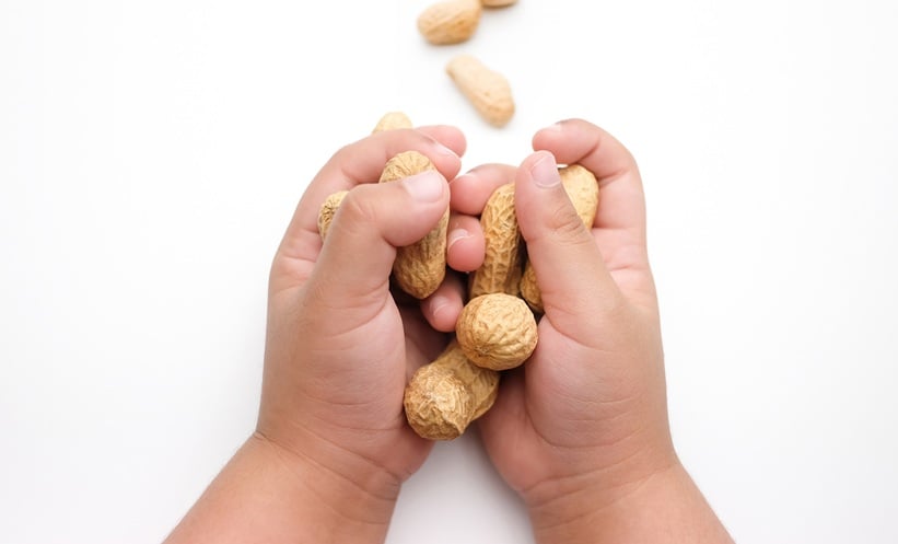
Childhood Food Allergy Risk Factors in Meta-Analysis
CHILDHOOD food allergy risk begins early, shaped by a complex interaction of genetic, environmental, and clinical factors, according to a large systematic review and meta-analysis. Analysing data from 190 studies involving approximately 2.8…
Continue Reading
-

Scientists Find Cancer Linked Chemicals In Nearly All Hair Extensions
Credit: Wikimedia Commons You’ll find glossy packets of hair extensions in most beauty supply stores. They promise a new look for a few dollars, but behind the glitter, scientists have found a cocktail of industrial chemicals…
Continue Reading
-
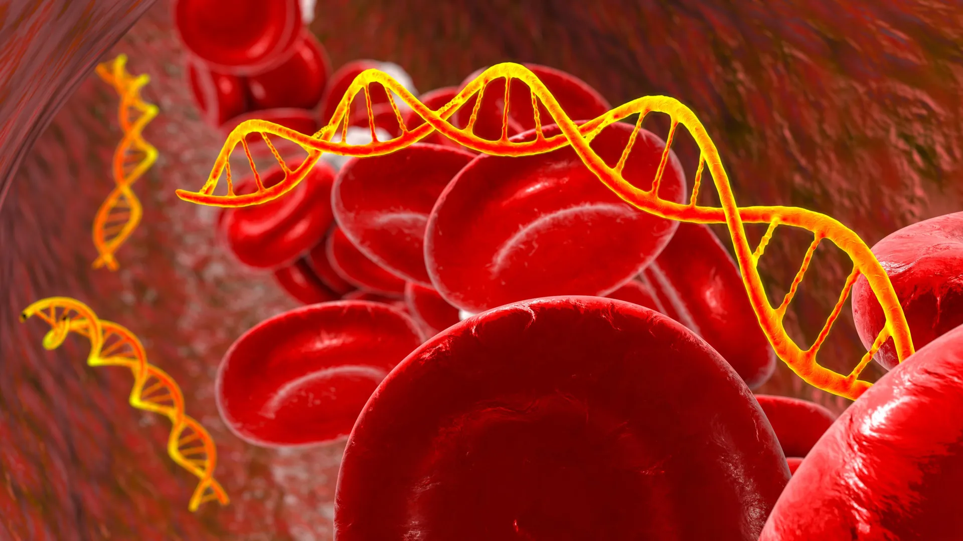
Mysterious RNA led scientists to a hidden layer of cancer
The journey began with T3p, a small RNA molecule detected in breast cancer but not in normal tissue. When it was first described in 2018, it stood out as unusual. That initial finding launched a six-year effort to systematically identify similar…
Continue Reading
-
catfish prevent disease spread, earthquake maps and student with math learning disabilities
The Science & Technology desk gathers a weekly digest of impactful and interesting research publications and developments at Stanford. Read the latest in this week’s Research Roundup.
Exploring African catfish as a sustainable…
Continue Reading
-
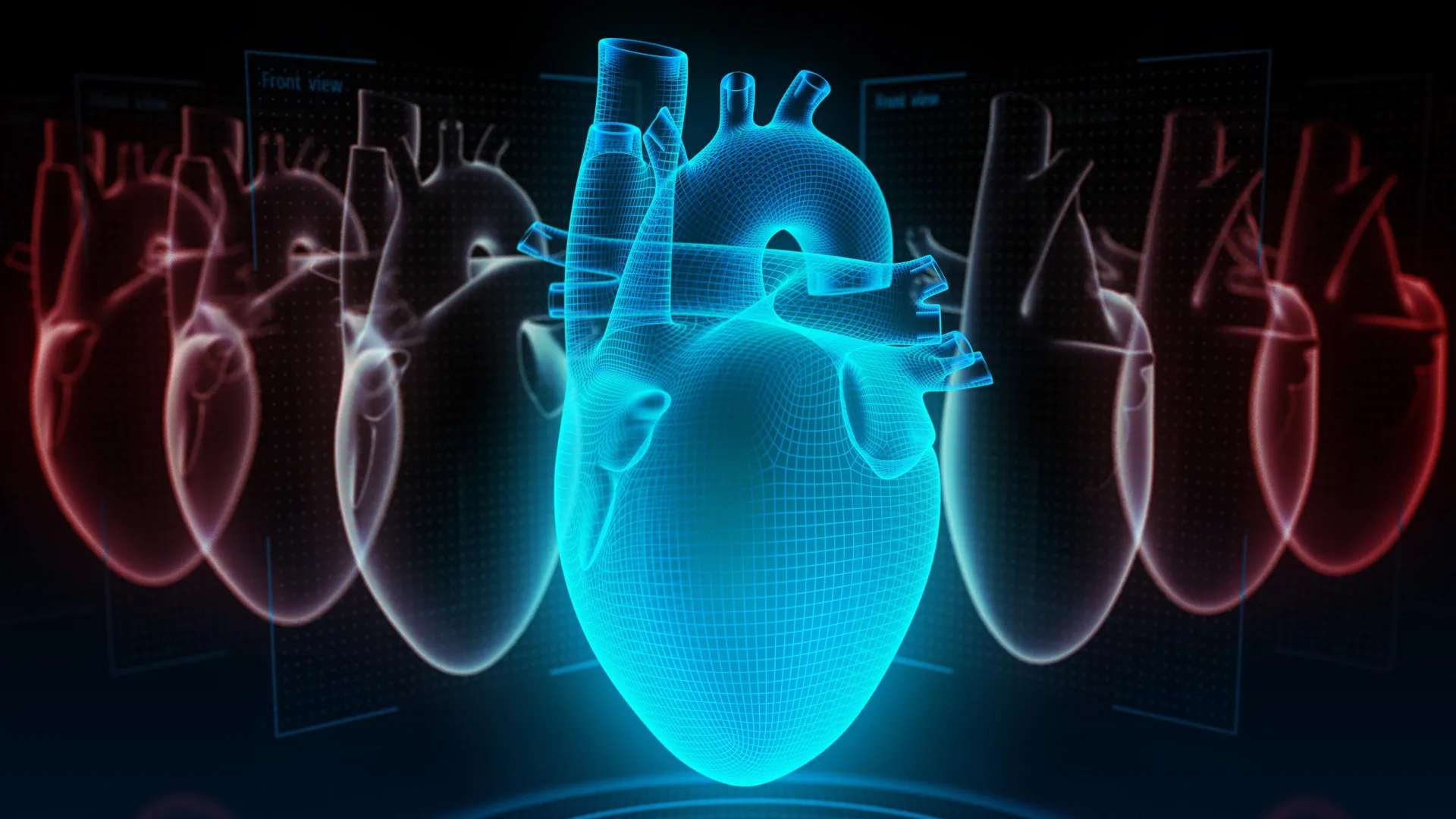
Nearly 200,000 people reveal the real key to heart health
A new study published in JACC, the flagship journal of the American College of Cardiology, suggests that the type of foods people choose on a low-carbohydrate or low-fat diet may be more important than simply cutting carbs or fat. Researchers…
Continue Reading
-
UMich researchers create digital AI copy of patient brain tumors
A University of Michigan study published Jan. 6 used machine learning to create a digital twin replica of gliomas to shed light on the metabolic pathways activated by brain tumors. Gliomas — tumors composed of the nervous system’s…
Continue Reading