At 4:30 a.m. you’ll find Kevin Hart in the gym.
While he’s best known for being a comedian and actor, Hart trains like a serious athlete: six days a week, about an hour and a half a day, no excuses.
It started over a…

At 4:30 a.m. you’ll find Kevin Hart in the gym.
While he’s best known for being a comedian and actor, Hart trains like a serious athlete: six days a week, about an hour and a half a day, no excuses.
It started over a…

Physicists have developed a new theory that brings together two major areas of modern quantum physics. The work explains how a single unusual particle behaves inside a crowded quantum environment known as a many-body system. In this setting, the…
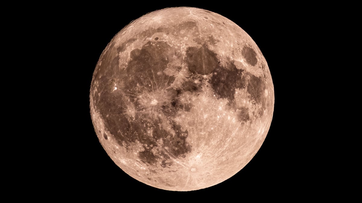
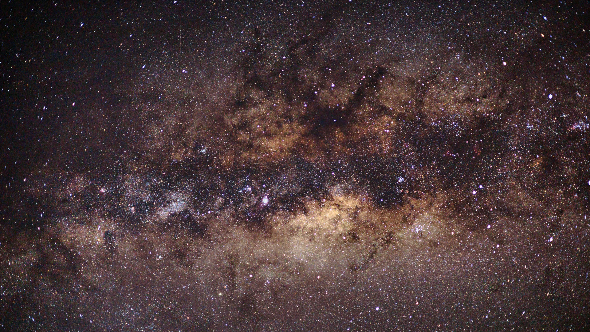
Tonight, the Moon looks slightly slimmer again as it approaches the Third Quarter. Its illuminated section is fading even more, meaning each night there’s less for us to see.
QUICK FACTS
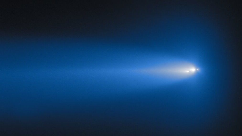
What it is: Comet C/2025 K1 (ATLAS)
Where it is: 220 million miles away, in the constellation Pisces
When it was shared: Jan. 28, 2026
Just as the mythological Icarus melted his feathers and wax wings when he flew too close to the sun,…
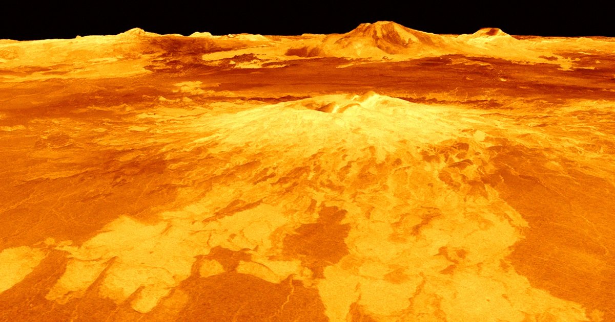
Venus has long been known as Earth’s evil twin. While they both are roughly the same size and formed in the same inner region of the solar system, Venus is far less hospitable to life as we know it. Its surface temperatures can reach over…
Astronomers have spent decades hunting for dark matter by looking for light that isn’t there — and that strategy has mostly failed.
Now, a team of researchers has tried something radically different. Instead of searching the sky, they…
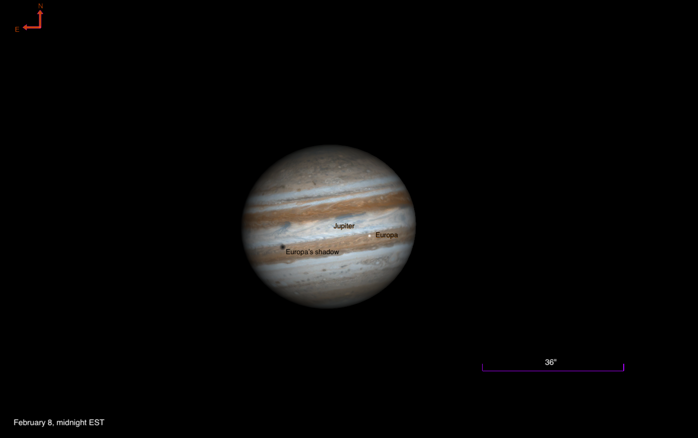
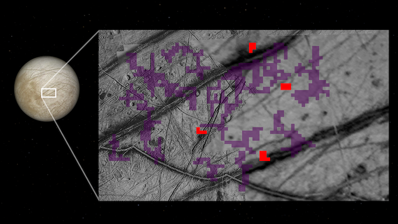
The icy moon Europa transits Jupiter tonight, followed by its shadow. Now past opposition, moons and their shadows are well separated.
Around midnight…

Beneath Earth’s surface lies a kingdom of undiscovered microscopic life. These “intraterrestrials” survive in some of the harshest conditions on the planet — and scientists are hunting for these microbes.
In this excerpt from “
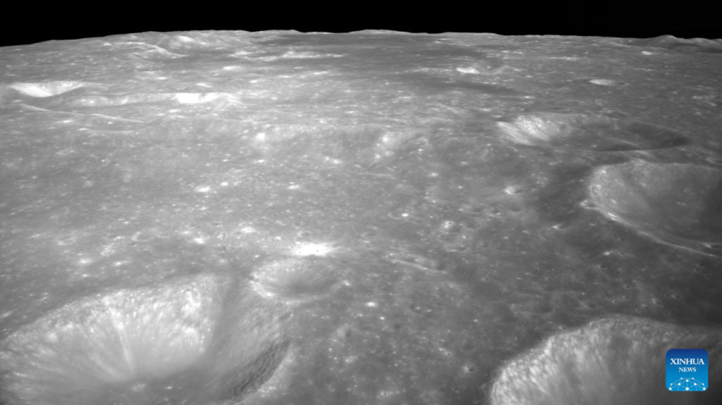
BEIJING – For the…