Qaasid News
Download Our App
Latest News from Pakistan
PhoneWorld Awards 2025: Celebrating the Best Smartphones Officially Available in Pakistan
December 31, 2025
Gold and silver see rollercoaster end to blockbuster year – BBC
December 31, 2025
Gold prices dip despite big jump in 2025 – Pakistan
December 31, 2025
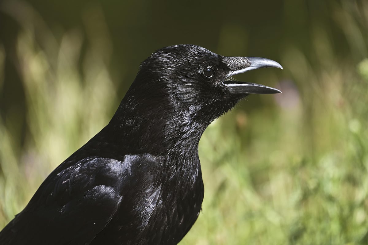
crows will come back for revenge after 17 years and will not forget
December 31, 2025
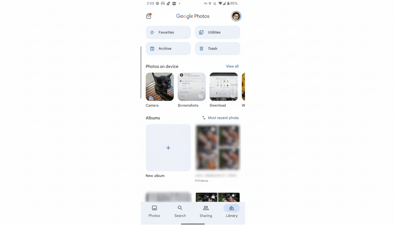
Google Photos Locked Folder Comes Under Fire
December 31, 2025
SAP announced final transition period for compatibility packs
December 31, 2025
NeuroVoices: David A. Hafler, MD, FANA, on Understanding MS as an Autoimmune and Neurodegenerative Disease | NeurologyLive
December 31, 2025
Cross-border rail passengers warned of new year disruption
December 31, 2025
‘They didn’t de-extinct anything’: can Colossal’s genetically engineered animals ever be the real thing? | Extinct wildlife
December 31, 2025
Travis Head wants rivals to share drinks after Sydney Test
December 31, 2025