Qaasid News
Download Our App
Latest News from Pakistan
More than 20% of videos shown to new YouTube users are ‘AI slop’, study finds | Artificial intelligence (AI)
December 27, 2025
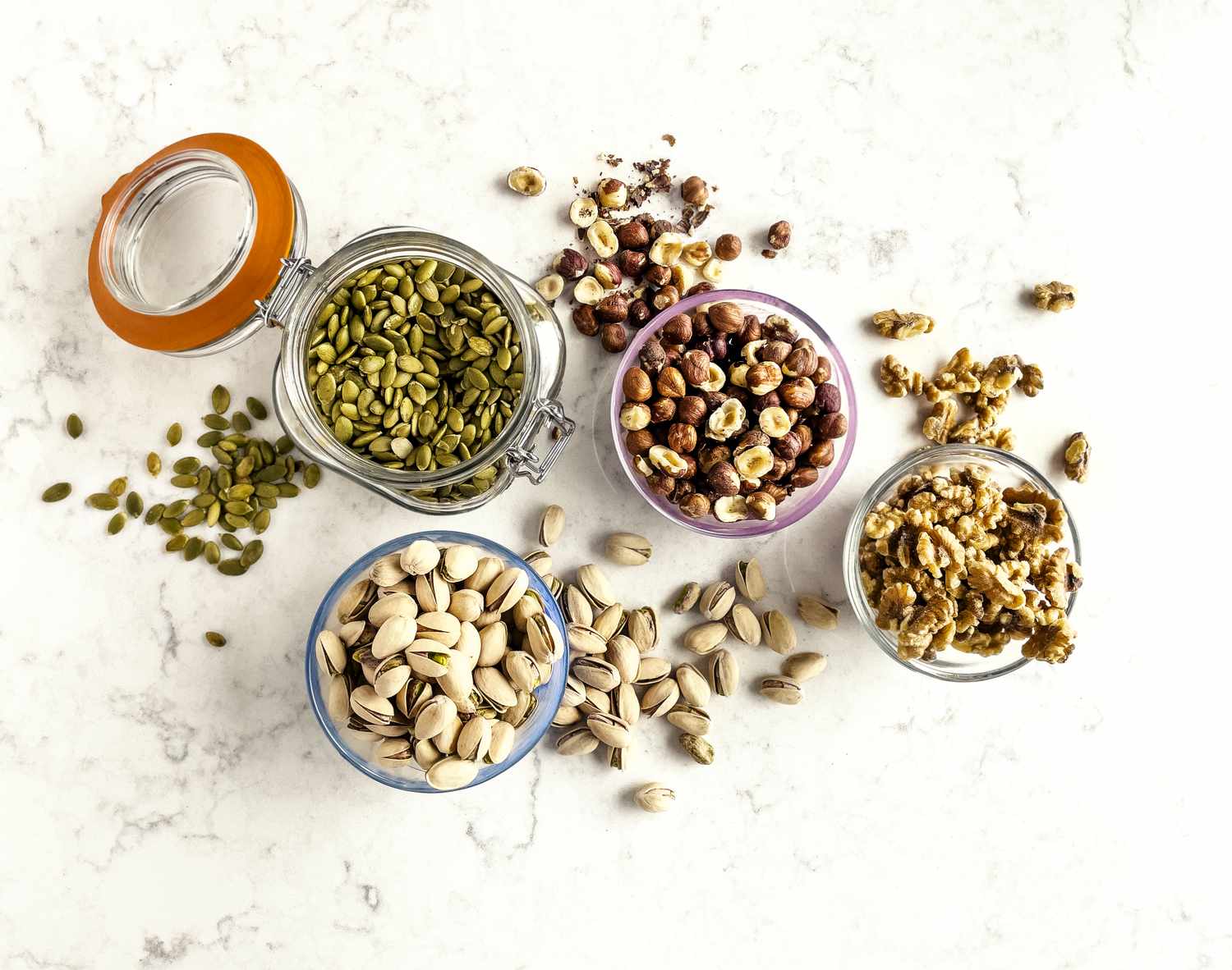
6 High-Protein Nuts and Seeds
December 27, 2025
Space.com headlines crossword quiz for week of Dec. 22, 2025: Which NASA observatory just completed its first sky map?
December 27, 2025
Malcolm's best tech in 2025: iPhone Air, Powerbeats Pro 2, and a cheap UPS – AppleInsider
December 27, 2025
Perspectives of Patients and Providers on Chronic Pain Assessment in Neurofibromatosis Type 1 (NF1): A Qualitative Study
December 27, 2025
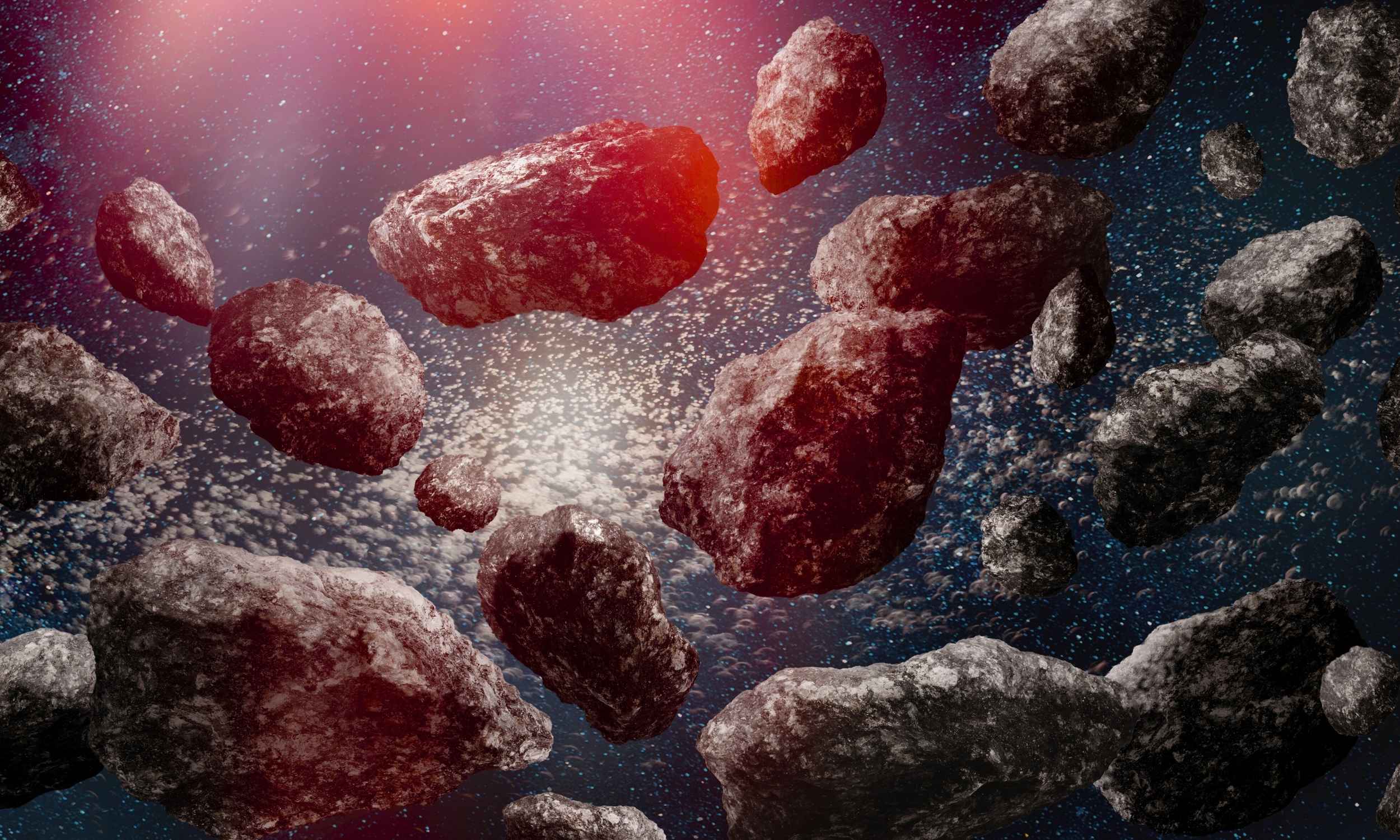
Astronomers find unexpected cluster of objects in the Kuiper Belt
December 27, 2025
Constructing A Science Of Stories
December 27, 2025
From Record Times to Postseason Qualifiers: A Midseason Look at UTRGV Swimming & Diving
December 27, 2025
Sophie Ellis Bextor reflects on life as mom of five
December 27, 2025
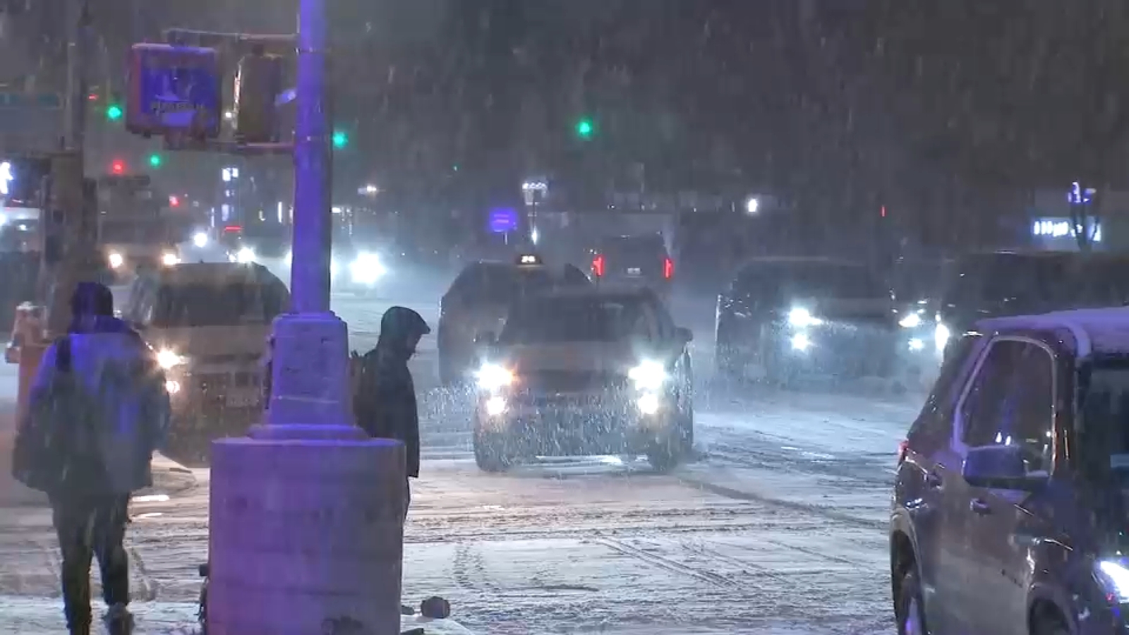
Winter storm NYC live updates: Snowstorm winding down across New York City, Tri-State area
December 27, 2025