Qaasid News
Download Our App
Latest News from Pakistan
PTI requests ECP to extend deadline for nomination papers in Islamabad local body polls – Dawn
December 25, 2025
Info Minister credits Quaid-e-Azam for Pakistan’s stature – RADIO PAKISTAN
December 25, 2025
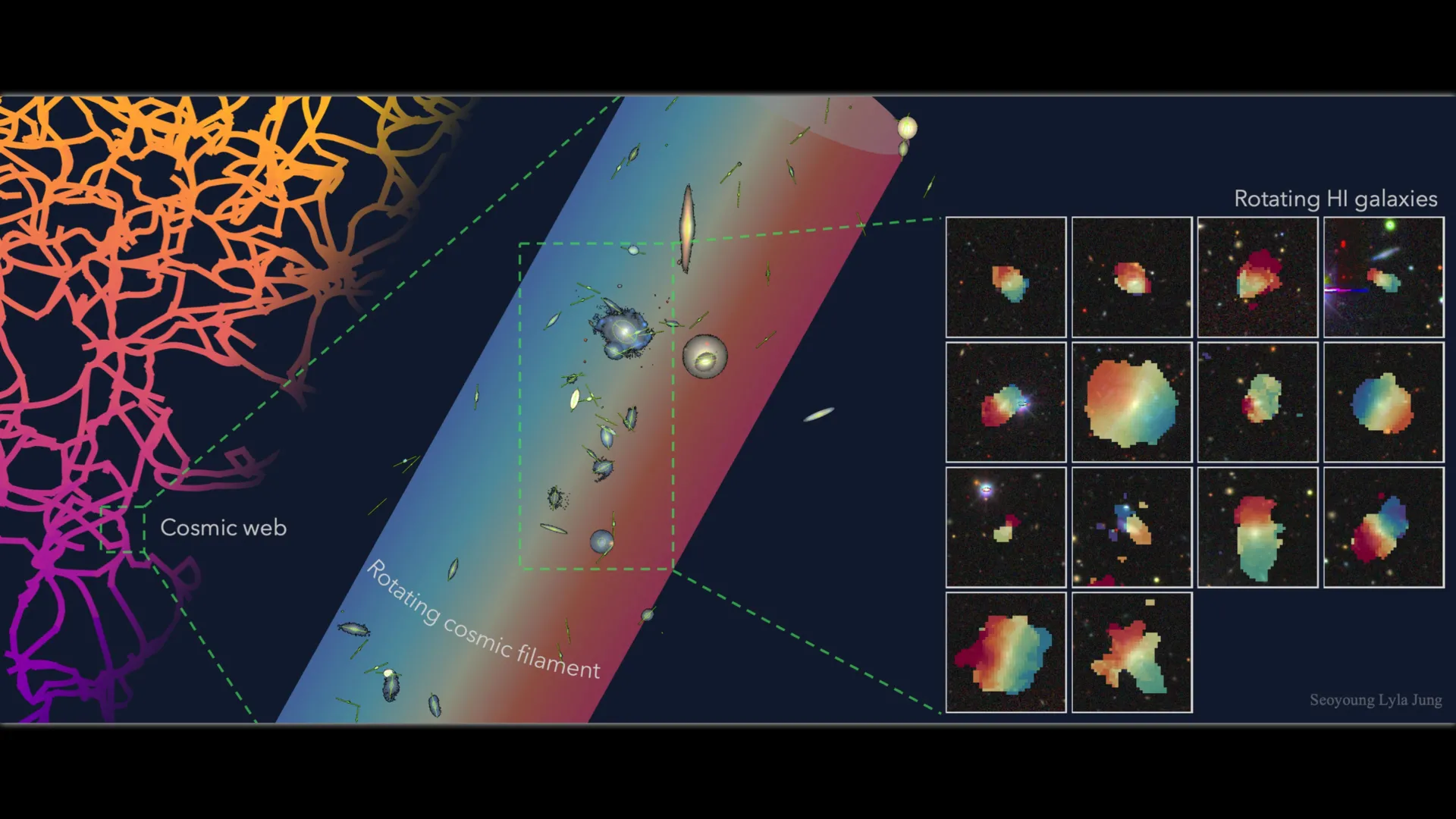
Astronomers discover one of the Universe’s largest spinning structures
December 25, 2025
‘Friends’ and ‘The Middle’ actor dies at 60
December 25, 2025
Thousands of jobs saved at North Sea oil and gas firm
December 25, 2025
Thousands of jobs saved at North Sea oil and gas firm – BBC
December 25, 2025
Pakistani official calls for media contribution to Upgraded Version 2.0 of CPEC-Xinhua
December 25, 2025
iPhone 18 Release Date: Is It Really Just Weeks Away, As New Report Claims? – Forbes
December 25, 2025
ITP issues traffic advisory due to Faizabad Loops extension work
December 25, 2025
Christian community celebrates Christmas across the globe – RADIO PAKISTAN
December 25, 2025