Qaasid News
Download Our App
Latest News from Pakistan
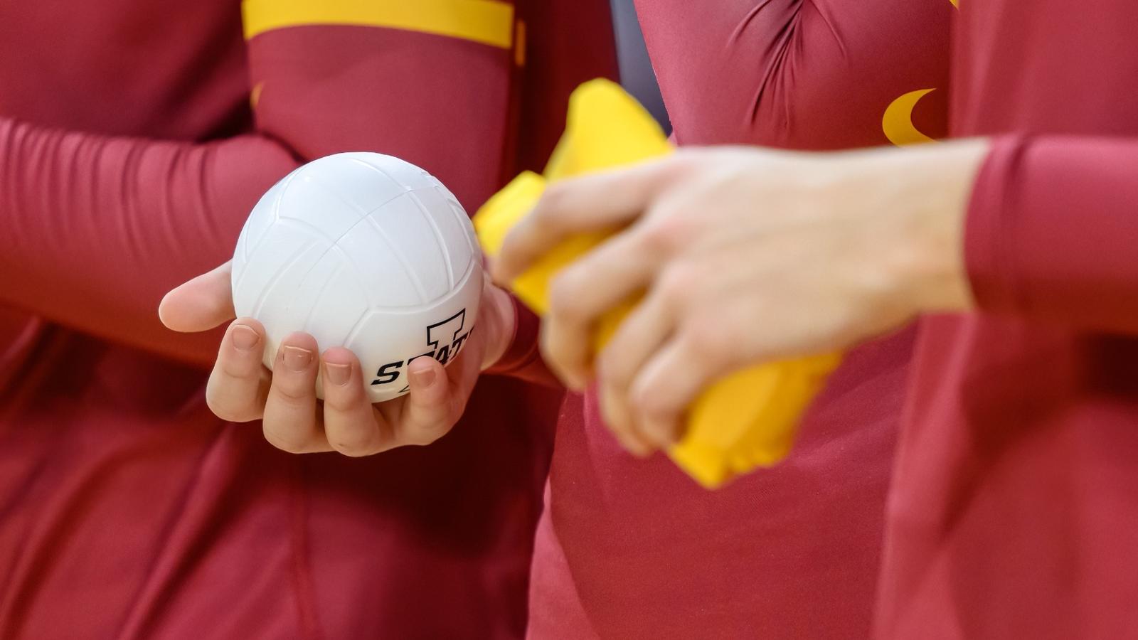
2026 Volleyball Camps Registration Now Open
December 20, 2025
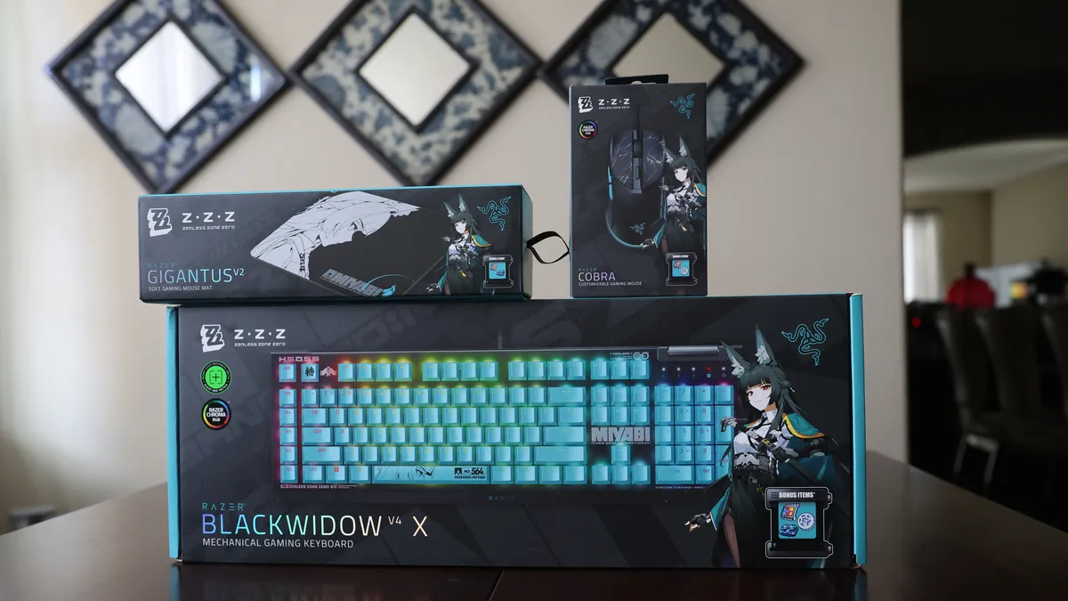
Hands-on with the Razer X Zenless Zone Zero PC gear
December 20, 2025
Minecoins, V-Bucks or Robux? How to give virtual cash to gamers | Christmas
December 20, 2025
Gymnastics coach from Maidstone returns to work after paralysis
December 20, 2025
New study traces how Earth’s alpine plant diversity evolved
December 20, 2025
This Week In Space podcast: Episode 190 — Holiday Special 2025
December 20, 2025
Galaxy S26 Ultra Release Date: Samsung Flagship’s Disappointing Delay – Forbes
December 20, 2025
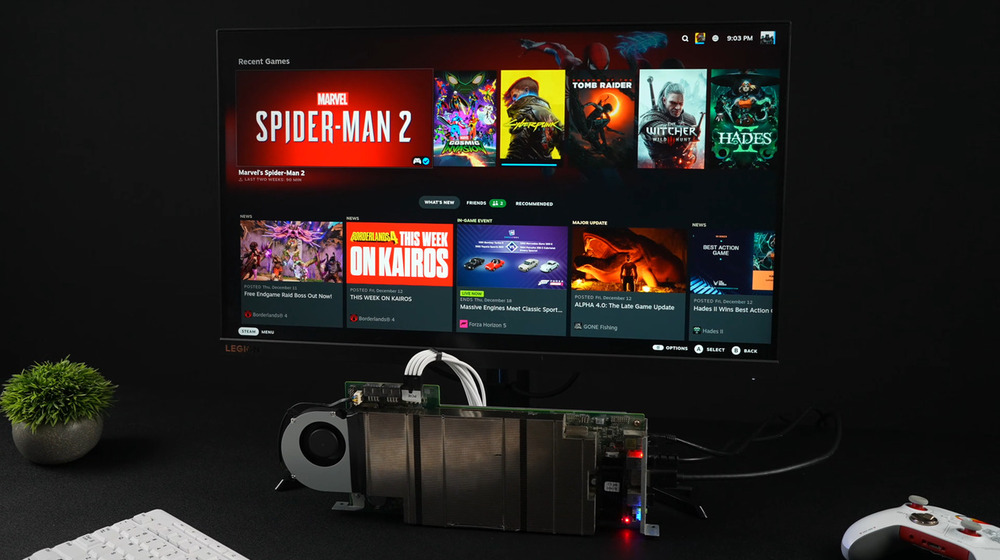
This DIY Steam Machine With Defective PS5 Chip Plays Spiderman 2 at 60 FPS
December 20, 2025
Minister of Foreign Affairs of Belarus M.Ryzhenkov meets management of National Museum of Oman – Министерство иностранных дел Республики Беларусь
December 20, 2025
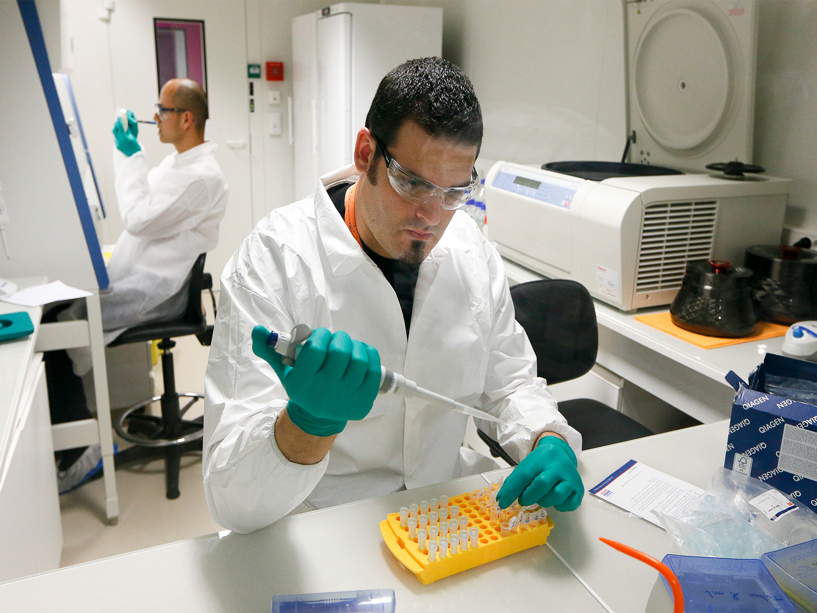
Pakistan poorly prepared to deal with HPAI threat, experts warn
December 20, 2025