
A new systematic evidence review finds that cannabis products that carry relatively high levels of the psychoactive compound tetrahydrocannabinol, commonly known as THC, may provide short-term improvements in pain and…
Category: 6. Health
-
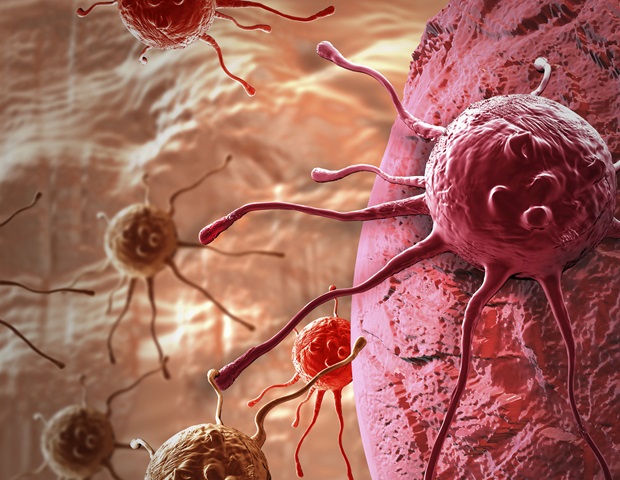
Reframing the role of MCL1 in cancer signaling and metabolism
A study by the Mildred Scheel Early Career Center group led by Dr. Mohamed Elgendy at the TUD Faculty of Medicine provides fundamental insights into cancer biology. Published in the renowned journal Nature Communications, the study…
Continue Reading
-
New Drug Stalls Alzheimer’s Development in Breakthrough Trial : ScienceAlert
Growing evidence suggests that the key to treating Alzheimer’s is catching it in its earliest stages. Now scientists have developed a promising new drug that seems to be effective at stalling the disease before it really gets started.
The…
Continue Reading
-
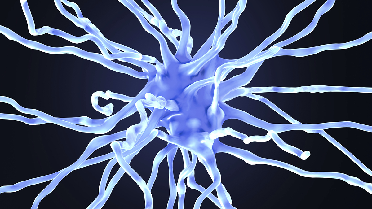
A senescence-based therapeutic approach for temporal lobe epilepsy
Temporal lobe epilepsy, which results in recurring seizures and cognitive dysfunction, is associated with premature aging of brain cells. A new study from researchers at Georgetown University Medical Center found that this form of…
Continue Reading
-

Research highlights a potential therapeutic strategy for vascular dementia
A possible new treatment for impaired brain blood flow and related dementias is on the horizon. Research by scientists at the University of Vermont Robert Larner, M.D. College of Medicine provides novel insights into the mechanisms…
Continue Reading
-
Whole-body imaging reveals where drugs bind at single-cell resolution
When you take a drug, where in your body does it actually go? For most medications, scientists can make only educated guesses about the answer to this question. Traditional methods can measure the concentration of a drug in an organ…
Continue Reading
-
Digital patient records improve survival in HIV treatment clinics
With 9.5% of its population estimated to be HIV-positive in 2019, Malawi has one of the highest rates of HIV prevalence in the world. While untreated HIV can lead to infection and death, antiretroviral therapy (ART)-a combination of…
Continue Reading
-
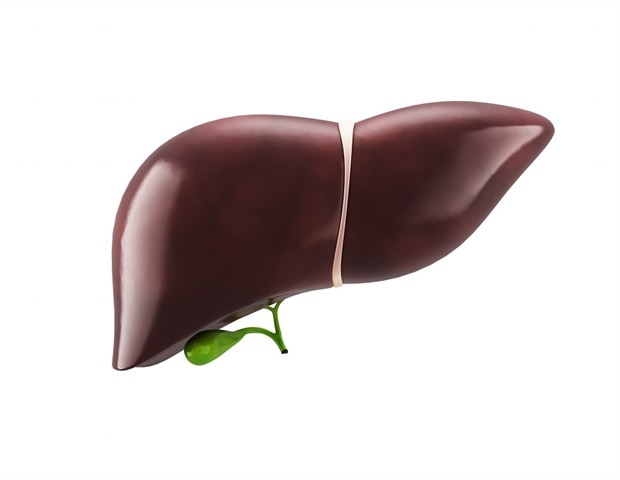
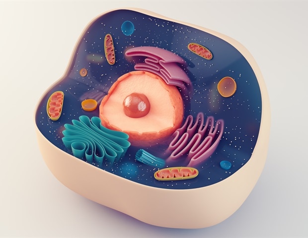
Fatty diets reprogram hepatocytes and increase liver cancer risk
One of the biggest risk factors for developing liver cancer is a high-fat diet. A new study from MIT reveals how a fatty diet rewires liver cells and makes them more prone to becoming cancerous.
The researchers found that in…
Continue Reading
-

Rethinking Mendelian assumptions in inherited retinal degenerations
A new study challenges what’s long been assumed about genetic variants thought to always cause inherited blindness. Investigators from Mass General Brigham used large public biobanks to determine that genes thought to…
Continue Reading
-

Early identification of nutrition risk in ICU patients using artificial intelligence
A new study by researchers at the Icahn School of Medicine at Mount Sinai suggests that artificial intelligence (AI) could help predict which critically ill patients on ventilators are at risk of underfeeding, potentially enabling…
Continue Reading