CEPI is the world’s largest funder of Nipah research and development. Its $150 million portfolio—including two Nipah vaccine candidates and a monoclonal antibody—spans the whole preparedness chain, from countermeasure development and…
Category: 6. Health
-

Efficacy and safety of clobazam adjunctive therapy in pediatric patien
Introduction
Pediatric drug-resistant epilepsy poses significant therapeutic challenges, with approximately 30%-40% of children failing to achieve sustained seizure free status despite the combined use of antiseizure medications (ASMs).1–3…
Continue Reading
-
Single enzyme mutation reveals a hidden trigger in dementia
Why do neurons die in dementia, and can this process be slowed down? A research group led by Prof. Marcus Conrad, Director of the Institute of Metabolism and Cell Death at Helmholtz Munich and Chair of Translational Redox Biology at the Technical…
Continue Reading
-
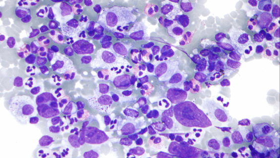
Israeli Lymphoma Treatment Reaches 100% Survival Rates in Large-Scale Study
Israeli researchers at a blood cancer conference in the US reported a lymphoma treatment that can achieve 100% survival rates. The results, compiled across 15 medical centers, revealed that patients treated with a combined…
Continue Reading
-

New therapy offers lifeline to ‘incurable’ blood cancer patients – The Times
- New therapy offers lifeline to ‘incurable’ blood cancer patients The Times
- Pioneering new treatment reverses incurable blood cancer in some patients BBC
- New study strengthens evidence for new CAR T-cell treatment for aggressive blood cancer
Continue Reading
-

Are pumpkin seeds really safe? Ayurveda expert warns too many may cause indigestion, weight gain, and low blood pressure
Pumpkin seeds have quietly earned a spot on the list of popular ‘superfoods’, thanks to their rich dose of magnesium, zinc, antioxidants, and healthy fats. Many people sprinkle them on salads, blend them into smoothies, or eat them as a…
Continue Reading
-
Hidden layers in the brain’s memory center revealed – Wiley Analytical Science
- Hidden layers in the brain’s memory center revealed Wiley Analytical Science
- Laminar organization of pyramidal neuron cell types defines distinct CA1 hippocampal subregions Nature
- Scientists find hidden layers in brain’s memory center
Continue Reading
-
Application Study of Health Management Model Based on Traditional Chin
Introduction
Chronic Obstructive Pulmonary Disease (COPD) is a chronic respiratory disease characterized by persistent airflow limitation, primarily caused by long-term exposure to harmful particles or gases such as smoking, air pollution, and…
Continue Reading
-
Characterization of drug resistance patterns, mutation profiles and pr
Introduction
Tuberculosis (TB), caused by the Mycobacterium tuberculosis complex, remains a major global health challenge and is now the leading cause of mortality from infectious diseases worldwide.1 According to the World Health Organization…
Continue Reading