Gerry Bradleyand
Auryn Cox,BBC News NI
 BBC
BBCThe principal of a County Londonderry school has said it is like “being back in Covid times”…

Gerry Bradleyand
Auryn Cox,BBC News NI
BBCThe principal of a County Londonderry school has said it is like “being back in Covid times”…
Friday, 5 December 2025, 17:57
The end of autumn and the beginning of winter in Malaga are both proving to be more complicated than in previous years due to the premature arrival of the flu season, which is currently four to…

MANHASSET, N.Y.–(BUSINESS WIRE)–Scientists at Northwell Health’s Feinstein Institutes for Medical Research have uncovered a critical mechanism driving vascular cognitive impairment (VCI), the world’s second most common form of dementia…

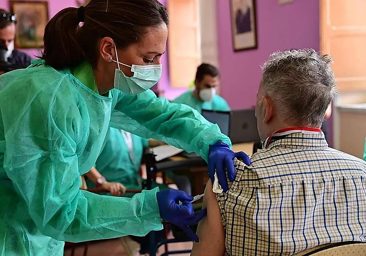
In an effort to reduce the ongoing Marburg Virus Disease (MVD) outbreak in Ethiopia, the Sabin Vaccine Institute has announced that it has sent about 640 doses of its investigational cAd3-Marburg Vaccine to Ethiopia.
As…
ADDIS ABABA, Dec. 5 (Xinhua) — Ethiopia’s Ministry of Health has announced that it is set to start administering Marburg virus vaccine to contain an outbreak of the disease in the southern part of Ethiopia.
The ministry, in a statement…
ADDIS ABABA, Dec. 5 (Xinhua) — Ethiopia’s Ministry of Health has announced that it is set to start administering Marburg virus vaccine to contain an outbreak of the disease in the southern part of Ethiopia.
The ministry, in a statement…

When the Centers for Disease Control and Prevention (CDC) altered its website last month to reflect US health secretary Robert F Kennedy Jr’s belief of a causal link between vaccines and autism – a claim that has been debunked by dozens of…

The US Centers for Disease Control and Prevention (CDC)’s vaccine advisers voted on Friday morning to limit hepatitis vaccines in a major move signaling the Trump administration’s regressive approach to vaccines that have been given safely…