An Oregon State University scientist and a team of undergraduate students have uncovered real-time insights into a chemical process linked with Alzheimer’s disease, paving the way toward better drug designs.
The researchers used a…

An Oregon State University scientist and a team of undergraduate students have uncovered real-time insights into a chemical process linked with Alzheimer’s disease, paving the way toward better drug designs.
The researchers used a…

FOOD ALLERGY (FA) risk was further clarified at the genetic level after a large genome-wide association meta-analysis identified 37 suggestive risk variants across children and adults.
FA is an immune-mediated condition, often driven by…
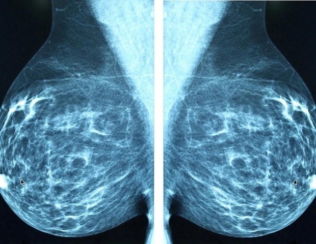
For many women with breast cancer, the very treatment that saves their lives can also bring fatigue, loss of muscle mass, emotional strain and other daunting obstacles. A new study shows that exercise during chemotherapy does more…
The risk of peanut allergy was significantly reduced if the younger siblings were introduced to peanuts in their first year of life
MILWAUKEE, Feb. 26, 2026 /PRNewswire/ — Younger…

A NEW cerebrospinal fluid (CSF) liquid biopsy approach could significantly improve how brain tumours are diagnosed, particularly in children, by using AI to analyse tiny amounts of tumour DNA found in cerebrospinal fluid. Researchers have…

A NEW retrospective study suggests that the red cell distribution width-to-platelet ratio (RPR) may help predict cardiac surgery-associated acute kidney injury (CSA-AKI), offering clinicians a potentially accessible biomarker for early risk…

ASTHMA remains a major cause of ill health in adults, and new research highlights how asthma symptoms are closely shaped by conditions inside the home. A large study of adults in Texas suggests that everyday indoor exposures, many of them…

SLEEP apnea specific hypoxic burden predicted higher 30-day postoperative mortality and cardiac complications after major surgery.
Obstructive sleep apnea is a common, heterogeneous condition…